Generic selectors
Exact matches only
Search in title
Search in content
Post Type Selectors
Filter by Categories
Brief Report
Case Report
Editorial
Focus
Images/Instrument in Dermatology/Dermatosurgery
Innovations
Letter to the Editor
Living Legends
Looking back in history
Original Article
Perspective
Resident Forum
Review Article
Spot the Diagnosis
Tropical Dermatology
Visual Treats in Dermatology
View/Download PDF

Translate this page into:

Visual Treats in Dermatology
2022
:2;
18
doi:
10.25259/CSDM_19_2022

Rough nails with reticulate oral lesions

Department of Dermatology and STD, Jawaharlal Institute of Postgraduate Medical Education and Research, Gorimedu, Puducherry, India
Corresponding author: Arunachalam Narayanan, Department of Dermatology and STD, Jawaharlal Institute of Postgraduate Medical Education and Research, Gorimedu, Puducherry, India.narayanan359@gmail.com
Licence
This is an open-access article distributed under the terms of the Creative Commons Attribution-Non Commercial-Share Alike 4.0 License, which allows others to remix, transform, and build upon the work non-commercially, as long as the author is credited and the new creations are licensed under the identical terms.

How to cite this article: Narayanan A, Ramar SY. Rough nails with reticulate oral lesions. CosmoDerma 2022;2:18.

A 35-year-old male patient presented with dystrophy of all 10 fingernails. The patient had brittle, thin nails with increased longitudinal ridging [Figure 1a] resulting in a rough, opaque appearance for 6 months. The patient had nontender, reticulate white lesions on his bilateral buccal mucosa [Figure 1b]. Based on the characteristic clinical appearance, we made a diagnosis of trachyonychia secondary to nail lichen planus (LP) with oral lichen planus. Trachyonychia is a disorder of the nail matrix seen in 10% of patients with nail LP. While nail LP is most often isolated in these patients, the most common type of LP associated with nail LP is oral LP.[1] Treatment is often challenging, and treatment options include topical clobetasol propionate, intramatricial triamcinolone, tazarotene gel, systemic retinoids, and cyclosporine.

Figure 1:
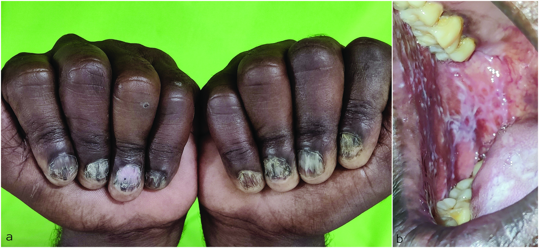
(a) Brittle, thin nails with increased longitudinal ridging resulting in a rough, opaque appearance (b) Reticulate, white lesions over the buccal mucosa.

Declaration of patient consent

The authors certify that they have obtained all appropriate patient consent.

Financial support and sponsorship

Nil.

Conflict of interest

There are no conflicts of interest.

References

  1. , , . Trachyonychia: A comprehensive review. Indian J Dermatol Venereol Leprol. 2011;77:640-5.
    [CrossRef] [PubMed] [Google Scholar]

Fulltext Views
4,307

PDF downloads
767
View/Download PDF
Download Citations
BibTeX
RIS
Show Sections