
Translate this page into:
Evolving role of lasers in nail therapeutics

*Corresponding author: Sachin Dhawan, Department of Dermatology, Fortis Memorial Research Institute, Gurgaon, Haryana and Skin n Smiles, Gurgaon, Haryana, Skin n Smiles, Gurgaon, Haryana, India. sac_dhawan77@yahoo.co.in
-
Received: ,
Accepted: ,
How to cite this article: Dhawan S, Sharma K. Evolving role of lasers in nail therapeutics. CosmoDerma 2022;2:19.
Abstract
The diseases of nails are chronic disorders due to the slow rate of growth of nails. The slow rate of nail growth results in long treatment regimens, thus having the potential of causing side effects and posing a limitation of administration to many people. Patients who suffer from organ dysfunction, elderly patients, patients under polypharmacy, and the inability of many patients to adhere to the complete regimen can cause the treatment of nail diseases to be frustrating for both the treating dermatologists as well as patients. Most nail disorders have formed a set treatment protocol for the administration of oral and topical drugs over the years. The use of lasers has yet to make its mark in the treatment of nail diseases due to lack of a universally accepted protocol. This review article looks into various studies evaluating the efficacy of lasers in nail diseases. Largely, this review is based on an evaluation of the effectiveness of lasers in onychomycosis (22 studies and 1 meta-analysis) and nail psoriasis (seven studies). While there is sufficient proof that lasers are effective in the treatment of nails, there is no gold standard for the type of lasers to be used for a particular disease, the treatment parameters, and the follow-up protocol. Evaluation of larger sample sizes against a control group and longer follow-ups are the need of the hour for the formulation of much-needed protocols.
Keywords
Lasers
Nail diseases
Onychomycosis
Nail psoriasis

INTRODUCTION
Studies discussing the potential use of lasers for nail disorders in clinical practice began to appear in the 1980s. These studies particularly focused on the high-powered carbon dioxide (CO2) lasers that were available at that time.[1,2] Mostly, these studies discussed the ablative effect of lasers in nail matrixectomies, their use in onychomycosis following total nail ablation, and for nail, fenestration to improve absorption of topical drugs.[1,3] The use of lasers for the treatment of nail abnormalities can be considered as a potential option to reduce the systemic side effects of oral treatments, as well as for patients who are unable to take these treatments due to preexisting organ dysfunction or drug interactions. With the increase in awareness among patients, there is an increase in the number of patients who want to proactively avoid long term oral medications after reading about the potential organ dysfunction on the internet. There have been some concerns over the years regarding the unproven efficacy of lasers, which also raises a concern for the investment costs involved in procuring the machines.[4] Over the years, many studies and article reviews comparing the efficacy of various lasers have been done, onychomycosis being one of the most extensively evaluated nail diseases.
The use of lasers in practice has been attractive for both doctors and patients alike for many reasons. The treatment duration of nail abnormalities usually extends over a long period of time. This poses difficulty to both the patients as well as the doctors. The biggest hurdle that remains is the adherence to treatment, as patients tend to either be irregular or gradually lose patience and trust over the treatment due to slow cosmetic improvement. The introduction of lasers has increased the possibility of shorter treatment regimens.
MATERIAL AND METHODS
This review article includes any original study, review article or analysis, published in a peer reviewed journal, that examined the use of laser technology in various nail abnormalities. The nail diseases were diagnosed microbiologically or histologically. Mycological cure (for onychomycosis), as well as clinical cure, were the parameters looked into, to measure the efficacy of laser treatment.
INCLUSION CRITERIA
An electronic database search was done on PubMed and ResearchGate to identify the papers which met the initial inclusion criteria. Searches were done using a combination of keywords “lasers,” “nails abnormalities,” “nail diseases,” “onychomycosis,” and “nail psoriasis.” No date limit was set by the authors, but as use of lasers in nails is not a very old modality, no study older than 2010 was included. Abstracts were reviewed to remove duplicates. Only papers that were written in English and had full study details were reviewed and included. The authors would have liked to have “duration of follow-up” as one of the inclusion criteria, but the lack of evidence and variable results with different durations of treatment prevented us from doing so. A total of 22 individual studies and 1 meta-analysis were evaluated for the treatment of onychomycosis and seven studies were evaluated for treatment of psoriasis by the authors.
ONYCHOMYCOSIS
Onychomycosis is a chronic fungal infection of the nail apparatus. It may involve the nail bed, nail plate, or matrix. It is difficult to treat and relapses are common, specifically due to the difficulty of adherence to treatment pertaining to its longevity. Onychomycosis is caused by dermatophytes Trichophyton rubrum most commonly, followed by Trichophyton mentagrophytes and Candida albicans.[5] The prevalence of onychomycosis is 2–13%. It can be up to 14–28% among elderly patients over 60 years of age.[6,7] Even after adequate oral antifungal treatment, recurrence (relapse or reinfection) is still common in about 10–53% of patients.[8] Topical drug treatments are not usually successful, as they cannot penetrate the nail plate.[9] Oral antifungal agents can have a significant risk of liver and kidney toxicity and drug interactions.[10] The limitations associated with oral antifungal treatment of onychomycosis have given rise to the need for a safer and an equally effective treatment option.
In 1984, Apfelberg presented the use of CO2 laser for onychomycosis. Since then, other laser treatments, such as long and short-pulsed 1064-nm Nd:YAG lasers, CO2 lasers, and lasers with wavelengths of 870 nm, 930 nm, and 1320 nm have been evaluated as potential new therapies for the treatment of onychomycosis.[1] A review article published by Bristow in 2014, evaluated 268 studies in which lasers were used for onychomycosis, out of which 12 eligible published studies were selected.[4] All 12 studies were published over the last 4 years. These studies included the use of long and short pulse 1064 nm Nd:YAG lasers as sole intervention, long pulse Nd:YAG laser in comparison to 1319 nm and broadband wavelength device, Q switched Nd:YAG 1064 nm/532 nm wavelengths system, 870/930 nm dual-band system or ablative carbon dioxide laser as a means to fractionate nails to enhance the penetration of topical antifungal agents by forming micro-channels[11-22] [Table 1]. Many studies excluded patients with severe or dystrophic onychomycosis. Out of these studies, only one paper offered a detailed design and protocol with a control group.[11] Most of the data published till this date has a low level of evidence due to small sample size, lack for randomized control groups for comparison of laser effectiveness, and lack of longer follow-ups to measure the rates of relapses and recurrence. Studies that did have longer follow-ups showed frequent relapses.[13,15] Various studies have successfully treated onychomycosis with long-pulse Nd:YAG laser in the energy range of 70-324 J with a pulse width of 30–45 ms and spot size of 4–6 mm.[22,26,30] Studies using Quasi long pulse (microsecond) Nd:YAG lasers have used fluence of 5-14 J/cm2 with a spot size of 2.5–6 mm.[15,17,19] A study with Q switched nanosecond Nd:YAG laser used fluence of 14 J/cm2 with a spot size of 5 mm, with sequential use of 1064 nm, followed by 532 nm wavelength [16] [Table 1]. Studies using fractional CO2 laser have used fluence range of 10-150 mJ.[18,24,31] In a study by Hees et al., in a 9-month follow-up, the authors noticed that while there was 65% mycological clearance within the first 6 months, the effect slightly reversed at the end of 9 months.[13] In a study by Hollmig et al., at 12 months there was no significant difference in clearance between laser treated and control group.[15] Therefore, the longevity of the effect of laser therapy demands more investigation. In a case report by Zawar et al. in 2017, the effectiveness of Q-switched Nd:YAG on a patient with recalcitrant onychomycosis was evaluated. The lateral and proximal nail folds were also treated. Fluence 500 mj and spot size 1.5 mm was used. Two passes with 1 min gap were done. There was no relapse at 3-month follow up.[23] A study by Arora et al. in 2019, assessed role of fractional CO2 laser with 1% terbinafine cream in 50 onychomycotic nails. After 4 monthly sessions, 88% nails showed culture clearance, and at 6-month follow-up, 88% of nails were culture-negative.[24] In a meta-analysis conducted by Ma et al. in 2019, a total of 35 articles involving 1723 patients and 4278 infected nails were included.[25] The evaluation of the studies in this analysis revealed that the overall mycological cure rate was 63.0%. The mycological cure rate of the long pulse 1064-nm Nd: YAG laser was 71.0%, of short pulse 1064-nm Nd:YAG laser was 21%, fractional CO2 laser was 45%, and of perforated CO2 laser was 95.0%. Hence, it was concluded that the efficacy of perforated CO2 laser treatment was higher than that of long-pulsed 1064-nm Nd:YAG laser. CO2 laser can both increase the localized temperature and gasify and decompose the infected tissue which has a sterilizing effect. Whereas, Nd:YAG laser only increases the nail temperature. In a study by Paasch et al., lasers of 808, 980, and 1064 nm were used to heat cell culture media and a nail clipping. The highest increase in temperature was found using a 980-nm laser with a pulse duration of 6 ms and a fluence of 27 J/cm2. The results for the 1064 nm system were almost comparable to 980 nm results. Thus, it was proven that complete fungal growth impairment can be achieved by raising temperatures above 50°C.[26] In a study by Wanitphakdeedecha et al., it was proved that 1064-nm long pulsed Nd:YAG laser inhibits the growth of the fungus. The long-pulsed 1064-nm Nd:YAG laser exhibited better efficacy than the short-pulsed (Q-switched) 1064-nm Nd:YAG laser. The cytoderm of Trichophyton fungi contains a large amount of melanin. Hence, the long-pulsed 1064-nm laser with a longer pulse width, leads to greater absorption of energy, giving rise to better therapeutic results.[27] The short-pulsed 1064-nm Nd:YAG laser leads to a comparatively lesser rise in temperature and mainly acts by producing sonic shock waves which inhibit the growth of the fungus.[13] The majority of patients in the included studies reported that they experienced a mild to moderate burning sensation during laser treatment. The combined efficacy of all laser treatments for onychomycosis in the analysis by Ma et al. was approximately 63%.[25] In comparison, the mycological efficacy of itraconazole pulse therapy and continuous terbinafine therapy for the treatment of onychomycosis were 79.6% and 84.8%, respectively.[28] The analysis by Ma et al. also showed the efficacy of CO2 perforated laser over that of fractional CO2 laser as 95% and 45% cure rates respectively due to photothermal effect of perforated CO2 laser. When compared to fractional CO2 laser, perforated CO2 laser produces a higher localized temperature. This can be difficult to control in terms of the depth of laser penetration, which can therefore result in larger wounds, forming a brown eschar, and having a higher risk of bleeding.[25] Yang et al. noticed that several patients felt mild transient pain with use of fractional CO2 laser, but the therapy showed a reliable efficacy at 1 and 3 months follow-ups for mild to moderate onychomycosis. On this basis, they concluded that the efficacy of fractional CO2 laser treatment could be improved safely by extending the duration of treatment (by increasing the time between two sessions).[29] In a study by Carney et al., it was shown that grinding of the affected nails before treatment helped in better penetration of laser. Thickness of less than 2 mm was found to be conducive to laser penetration.[12] Chen et al. found that application of 5% salicylic acid for 48 hours before using long-pulsed Nd:YAG laser enhanced clinical cure rates to 71% as compared to 49% achieved with only laser treatment.[33] Landsman et al. used 870-nm and 930-nm lasers to treat severe onychomycosis and this produced a mycological cure rate of only 38%.[11] Use of 1320-nm Nd:YAG laser by Ortiz et al. reported a lower curative efficacy than the control group.[30] A two-stage study involving 22 patients was published by Shan Zhong et al. in 2019. Patients were treated with a long-pulsed Nd:YAG 1064-nm laser. The first stage was performed once a week for 8 weeks, and the second stage was done once every 4 weeks for four visits. The mycological clearance rate and the clinical efficacy rate of the nails were 29% and 21% after the first stage, 69% and 35% after the second stage, and 67% and 39% during follow-up, respectively. This study demonstrated that the efficacy of the treatment significantly improved after the second stage of treatment as compared to the first stage, suggesting that the second phase and a longer follow up period were necessary.[31] Studies have also proved a better mycological and clinical efficacy of laser treatment combined with topical drugs due to the formation of microchannels, than that produced by laser treatment alone. In a study by Zhou et al., the mycological cure for laser vs. laser plus drug group was 39% and 57% respectively, and clinical cure was 53% and 73% respectively.[32] In a study by Chen et al., the mycological cure for laser vs. laser plus drug group was 72% and 82% respectively, and clinical cure was 49% and 71%, respectively.[33]
| Authors | Study type | No. of patients (nails) | Laser used | Sessions and intervals | Follow-up period | Parameters used | Study endpoint |
|---|---|---|---|---|---|---|---|
| Landsman et al.[11] | Randomized control trial | 36 (53) | 870-930 nm laser (with sham control) | 4 sessions in 2 months (day 1, 14, 42 and 120) | 9 months | Energy 424 J/cm2, spot size 1.5cm | Decrease in affected nail area and negative culture |
| Carney et al.[12] | Case series | 10 (18) | 1064 nm quasi long pulse Nd: YAG laser | 4 sessions (at 1, 2, 3 and 7 weeks) | 6 months | Energy 16 J/cm2, spot size 5 mm, pulse duration 300 µs, frequency 2 Hz | No significant decrease in area of nail involvement with Onychomicosis Severity Index (OSI) and negative cultures |
| Hees et al.[13] | Comparative study | 10 (20) |
1064 nm Nd: YAG. Long pulse (left toe nail) VS. Short pulse (right toe nails) |
2 sessions, 4 weeks apart | 9 months |
Energy 50 J/cm2, spot size, 3mm, pulse duration 40 ms Energy 25 J/cm2, spot size 1.5 mm, pulse duration 100 µs |
Decrease in area of nail involvement with OSI and negative histology and cultures in first 6 months and slight reversal at the end of 9 months. |
| Hochman[14] | Case series | 8 (12) | 1064 nm short pulse Nd: YAG laser with topical anti-fungal | 2 or 3 sessions, 3 weeks apart | 6 months after final treatment |
Energy 223 J/cm2, 0.65ms pulse width, spot size 2 mm 2 passes |
Significant visual improvement (not quantified) and negative fungal cultures. |
| Hollmig et al.[15] | Randomized control trial | 27 (125) | 1064 nm Quasi long pulse Nd: YAG laser | 2 sessions, 2 weeks apart | 12 months | Energy 5 J/cm2, spot size 6mm, pulse width 300 µs, frequency 6 Hz | Negative cultures and clearance at 3 months (33% patients). No difference at 12 months in treated and control group. |
| Kalokasidis et al.[16] | Self-control study | 131 | Nail reduction with drill and 1064 nm/532 nm Q Sw Nd: YAG laser | 2 sessions, 1 month apart (both 1064 nm and 532 m) | 3 months |
Fluence 14 J/cm2 Spot size 2.5mm Pulse duration 9nsec Frequency 5 Hz. 1064 nm followed by 532 nm. |
Decrease in area of nail involvement with OSI and negative cultures (95.4%) |
| Kimura et al.[17] | Self-control study | 13 (37) | 1064 nm quasi long pulse Nd: YAG laser | 2-3 sessions, 1 to 2 months apart | 6 months | Energy 14 J/cm2, spot size 5mm, pulse width 300 µs, frequency 5 Hz | Improvement of nail turbidity score and negative culture (51% nails) at 4 months |
| Lim et al.[18] | Case series | 24 | Fractional CO2 laser | 3 sessions, 1 month apart (with topical anti-fungal) | 6 months | Energy 160 mJ, density 150 spots/cm2, 2 to 3 passes | Decrease in affected nail surface area (92%) and negative microscopy (50%) |
| Moon et al.[19] | Case series | 13 (43) | 1064 nm Quasi long pulse Nd: YAG laser | 5 sessions, 1 month apart | 6 months |
Fluence 5J/cm2 Pulse duration 300 µs Frequency 5 Hz, Spot size 6mm |
Decrease in affected nail surface area and negative microscopy |
| Noguichi et al.[20] | Case series | 12 (12) | 1064 nm Quasi long pulse Nd: YAG laser | 3 sessions, 1 month apart | 3 months and 6 months | Energy 10 J/cm2, spot size 6 mm, pulse duration 500 µs (4 passes) | Improvement in 25% affected nails showing significant decrease in surface area |
| Waibel et al.[21] | Comparative study |
21 (21); 7 in each group |
1064 nm Nd: YAG vs. 1319 nm vs. broadband light |
4 sessions, 1 week apart | 1, 3 and 6 months | Clinical clearance. 100% clearance with 1064 nm and broadband light. 1 failure with 1319 nm laser. | |
| Zhang et al.[22] | Comparative study | 33 (154) | 1064 nm long pulse Nd: YAG laser | 4 OR 8 sessions, 1 week apart | 6 months | 240-324 J/cm2, pulse duration 30 ms, spot size 3 mm, frequency 1 Hz | No significant difference in mycological (51% & 53%) cure rates. Recurrence in 5 patients (10 nails) after 2-4 months after study. |
| Zawar et al.[23] | Case report | 1 (1) | 1064 nm QS Nd: YAG laser | 3 sessions, 2 weeks apart | 3 months | Fluence 500 mj, spot size 1.5mm. 2 passes with 1 min gap | Clinical and mycological clearance |
| Arora et al.[24] | Observational study | (50) | Fractional CO2 laser | 4 sessions 1 month apart | 6 months after last session | Energy 110 mJ, 256 spots/cm2, pulse interval 0.5 mm, pulse duration 0.1 ms. | 90% KOH and 88% culture negativity after 4 sessions, 86% KOH and 88% culture negativity after 6 months |
| Wanitphakdeedecha et al.[27] | Self-control study | 35 (64) | 1064 nm long pulse Nd: YAG laser |
4 sessions, 1 week apart (repeat cycle after 1 month if microscopy positive) |
At 3 and 6 months | Energy 35-45 J/cm2, spot size 4 mm, pulse width 30-35 ms, frequency 1 Hz | Cure rate 63.5%, 57.7% and 51.9% at 1, 3 and 6 month follow up |
| Yang et al.[29] | Self-control study | 18 (71) | Ultrapulse CO2 fractional laser | 4 weekly sessions, then one session every 2 weeks for 8 weeks | At 1 and 3 months | Energy 5 J/cm2, spot size 3 mm | 61.97% mycological cure at the end of 3 months |
| Oritz et al.[30] | Randomized control trial | 10 (20) | 1320 nm Nd: YAG laser | 4 sessions (day 1, 7, 14 and 60) | After 3 months | Spot size 5 mm, pulse width 350 ms, frequency 20 Hz | 50% mycological cure |
| Zhong et al.[31] | Self-control study | 22 (100) | 1064 nm long pulse Nd: YAG laser | 8 weekly sessions, then 1 session every month for 4 months | 3 months after last treatment | Energy 35-40 J/cm2, pulse duration 35 ms, spot size 4mm, frequency 1 Hz | Mycological cure (67%) and clinical cure (39%) at follow up |
| Zhou et al.[32] | Randomized control trial | 60 (223) |
Fractional CO2 laser with 1% luliconazole AND Fractional CO2 laser alone |
1 session every 2 weeks for 6 months with or without daily topical application | 6 months | Energy 10-15mJ, spot size 4-10 mm, pulse duration 0.5-1.0 seconds |
Mycological and clinical cure respectively; Laser: 39% and 53% Laser plus topical: 57% and 73% |
| Chen et al.[33] | Comparative study | 66 (140) |
1064 nm long pulse Nd: YAG laser AND Salicylic acid for 48 hours followed by laser |
Weekly for 4 weeks, then monthly for 6 months | 9 months | Energy 70-100 J/cm2, pulse width 45 ms, spot size 3 mm |
Mycological and clinical cure respectively; Laser: 72% and 49% Laser plus topical: 82% and 71% |
| Myers et al.[35] | Self-control study | 12 | 1534 nm long pulsed erbium glass laser | 3 monthly sessions | 7 months | Energy 100 mJ, pulse duration (3 ms long pulse and 6 ns short pulse), spot size 2mm | Clinical clearance with new nail growth |
| Zhang et al.[36] | Intra-patient comparative study |
9 (20+20) Bilateral involvement |
2940 nm Er:YAG fractional laser with 5% amorolfine lacquer AND 5% amorolfine lacquer only |
6 sessions (at 1, 2, 3, 4, 8 and 12 weeks) with twice weekly application of lacquer | 6 months | Energy 35-62 J/cm2, spot size 483 microns, density 120 spots/square | 90% clinical cure and 75% mycological cure in group 1. |
In a study by Belikov et al., the use of Ytterbium sensitized Erbium glass laser for active drug delivery in onychomycosis was demonstrated with healthy nail plates of seven patients (250 samples). Pulse duration of 270 μs, energy 4 mJ, spot size 220 μm, and 30 Hz frequency were used.[34] In a study by Myers et al., 12 patients were successfully treated with a long-pulsed erbium glass 1534 nm laser.[35] Zhang et al. studied the effect of combination of fractional 2940 nm Er:YAG laser with 5% amorolfine lacquer in onychomycosis. The study proved enhanced penetration of topical antifungal drugs due to microchannels created by the laser.[36]
Conclusion: It is clear that lasers have a definite role to play in the treatment of onychomycosis, especially in certain patient populations with systemic disease and the elderly. At present, non-thermal laser therapy (635 nm/405 nm dual diode laser) is the only FDA approved for the treatment of onychomycosis.[37] The analysis done by the authors infers that perforated CO2 laser has the best outcome, followed by long-pulsed Nd:YAG laser. Fractional CO2 lasers and short-pulsed 1064 nm Qs Nd:YAG lasers have lower cure rates, but in conjunction with topical antifungals (for fractional CO2) and with the use of quasi long pulse (300 μs) mode of Nd:YAG laser, they can shorten the length of treatment and improve the outcomes, since they are the more readily available technologies in dermatology clinics. Other fractional lasers like Er:YAG and Er:Glass laser can be used in place of fractional CO2 laser, based on availability. (Figure 1, 2 and 3 share the experience of authors in role of Qs Nd:YAG laser combined with fractional CO2 laser for treatment of onychomycosis.)

- A 54 year old female presented with a clinical diagnosis of onychomycosis of both great toes since more than two years, which showed no improvement after one year of oral antifungals.
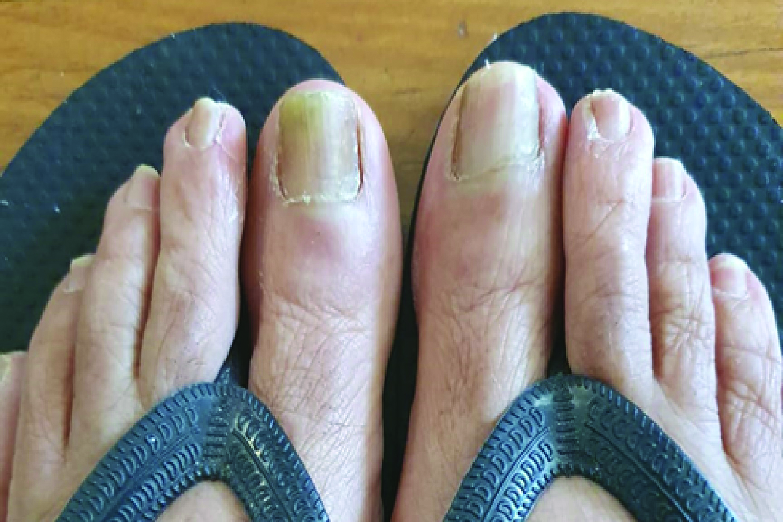
- She underwent 3 monthly sessions of Q switched Nd:YAG laser with 3 passes at wavelength 1064 nm in nanosecond mode with spot size 6 mm and energy 4 J/cm2, followed by 6 passes with spot size 8 mm and energy 2.2 J/cm2. Fractional CO2 laser was then done with 100 micro beams at 30 mJ energy. Follow-up after 3 months from last sessions showed marked clinical improvement.
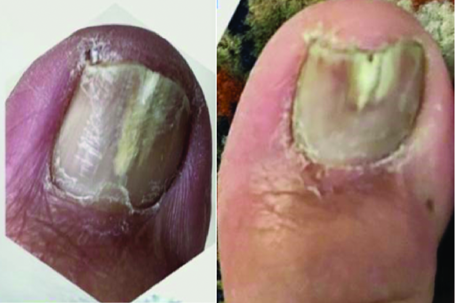
- A 60 year old male presented with a clinical diagnosis of onychomycosis of right great toe, which showed no improvement after six months of oral antifungals. He was given 3 monthly sessions of Q switched Nd:YAG laser with 2 to 3 passes at wavelength 1064 nm in nanosecond mode with spot size 6mm and energy 4 J/cm2, followed by 5 passes with spot size 8 mm and energy 2.2 J/cm2. Fractional CO2 laser was then done with 100 micro beams at 30 mJ energy. Follow-up after 3 months from last sessions showed clinical improvement as reduction in the area of involvement.
NAIL PSORIASIS
In patients suffering from chronic plaque psoriasis, the prevalence of nail psoriasis documented in the literature is over 50%, with an estimated lifetime incidence of 80–90%.[38] The cosmetic handicap in nail psoriasis is sometimes so extensive that patients tend to hide their hands and/or feet and shy away from social interactions. Hence the involvement of nails in psoriasis may have a substantial negative impact on the psychological, physical, and social aspects of the life of an individual.
Treatment of nail psoriasis poses a great difficulty due to slower improvement rates. This is due to slow rate of growth, poor drug delivery due to less absorption through nail plate and hyperkeratosis of affected nails, side effects of the systemic treatment options, and difficulty in adherence to treatment due to prolonged treatment regimens.[39]
As angiogenesis is the main pathology in nail psoriasis, most studies on lasers for nail psoriasis have evaluated the use of pulsed dye laser (PDL). PDL specifically targets blood vessels. In a study by Oram et al., 595-nm PDL was used. It showed 86% improvement at 1 month, mainly in the nail bed nail psoriasis severity index (NAPSI) score.[40] Another study by Treewittayapoom et al., compared two different pulse widths. The pain was significantly more intense in the group with longer pulse, but no difference was observed in the treatment outcome between the long 6 ms pulse duration with 9 J/cm² energy group and the short 0.45 ms pulse duration with 6 J/cm² energy group. A rapid decrease in NAPSI, followed by a significant increase after the third month of treatment despite ongoing treatment was reported, thus giving rather contradictory results.[41] However, in a study by Huang et al., a significantly higher percentage of patients had improvement after 6 months of treatment with PDL plus topical tazarotene than after tazarotene treatment alone.[42] In a study by Al-Mutairi et al., a comparison of the PDL with the excimer laser was done. A total of 304 nails, 148 with excimer laser, and 156 with PDL, were treated. Complete nail recovery was shown in 14% of hands treated with PDL at week 12, while no hands achieved NAPSI-75 at week 12 with an excimer laser. This proved the effectiveness of PDL over excimer laser. Subungual hyperkeratosis and onycholysis improved significantly, while nail pitting was the least responsive. Oil drops and splinter hemorrhages showed moderate response.[43] Another study by Arango-Duque et al. compared PDL with long-pulsed 1064 nm Nd:YAG laser. Both groups were treated with calcipotriol betamethasone gel. All patients showed improvement in nail bed and nail matrix psoriasis with no statistical difference between the results of the two lasers, though the administration of long-pulsed 1064 nm Nd:YAG was more painful.[44] In a study by Kartal et al., at the end of three treatment sessions done 1 month apart, both nail bed and matrix lesions significantly responded to long-pulsed 1064 nm Nd:YAG laser treatment.[45] A study by Khashaba et al. evaluated the efficacy and safety of long-pulsed 1064 nm Nd: YAG laser for treatment of nail psoriasis. The study included 22 patients with bilateral fingernail psoriasis. They were randomly assigned to either four sessions of long-pulsed Nd:YAG laser once monthly, or daily topical placebo for 4 months, followed by 3 months follow-up. The evaluation was done using NAPSI score at baseline, second month, fourth month, and after follow-up period. There was a statistically significant improvement in NAPSI score as well as dermoscopic findings in the nails treated by laser. Nail bed showed better improvement than nail matrix.[46] [Table 2] gives a brief overview of the various studies that were evaluated by the authors.
| Authors | Study type | No. of patients (nails) | Laser used | Sessions and intervals | Follow-up period | Parameters used | Study endpoint |
|---|---|---|---|---|---|---|---|
| Authors | Study type | No. of patients (nails) | Laser used | Sessions and intervals | Follow-up period | Parameters used | Study endpoint |
| Authors | Study type | No. of patients (nails) | Laser used | Sessions and intervals | Follow-up period | Parameters used | Study endpoint |
| Oram et al.[40] | Case series | 5 | Pulsed dye laser (595 nm) | 3 sessions, 1 month apart |
Pulse duration 1.5 ms, Spot size 7 mm, Energy 8 to 10 j/cm2 |
Significant reduction in nail psoriatic severity index (NAPSI) score | |
| Treewittayapoom et al.[41] | Comparative study | 20 (79) | Pulsed dye laser (595 nm) | 1 session | 6 months | 6 ms pulse duration with 9 j/cm2 OR 0.45 ms pulse duration with 6 j/cm2 | Reduction in NAPSI score, no significance difference in both groups |
| Huang et al.[42] | Comparative intrapatient (left to right control) study | 25 | Pulsed dye laser (595 nm) with 0.1% tazarotene cream OR only 0.1% tazarotene cream | 6 sessions, 1 month apart | 6 months |
Pulse duration 1.5 ms, Beam diameter 7mm, Energy 9 J/cm2 |
Reduction in NAPSI score, significantly more in group with PDL |
| Al-Mutairi et al.[43] | Comparative intrapatient (left to right control) study |
42 (304) (148) (156) |
Pulsed dye laser (595 nm) 308 nm excimer |
3 sessions, 1 month apart Twice weekly sessions for 3 months |
6 months |
Pulse duration 1.5 ms, beam diameter 7 mm, energy 8 to 10 j/cm2 Minimal erythema dose calculated over unaffected skin and fluence was increased by 200 mJ every session (maximum 5000 mJ/cm2 |
NAPSI improvement was significantly greater in PDL than excimer laser. |
| Arango-Duque et al.[44] | Prospective intra-patient (left to right controlled) study |
13 Right hand Left hand |
Pulsed dye laser (595 nm) 1064 nm Nd: YAG laser |
4 monthly sessions (with application of betamethasone calcipotriol gel every day for 1 week for both groups) |
4 months |
Energy 6 J/cm2, spot size 7 mm, pulse duration 0.4 ms Energy 40 J/cm2, spot size 5 mm, pulse duration 35 ms. |
Reduction in NAPSI score, no significance difference in both groups |
| Kartal et al.[45] | Case series | 16 | 1064 nm long pulse Nd: YAG laser | 3 sessions, 1 month apart | 3 months |
Pulse duration 15 ms, Spot size 6mm, Energy 10 J/cm2 Frequency 1.5 Hz |
Significant reduction in NAPSI |
| Khashaba et al.[46] | prospective intra-patient left-to-right, randomized, placebo-controlled study | 22 |
1064 nm long pulse Nd: YAG laser OR Topical placebo |
4 sessions, 1 month apart | 4 months treatment followed by 3 months follow up |
Pulse duration 35 ms, Spot size 5mm, Energy 40 J/cm2 |
Significant reduction in NAPSI on the laser side |
Conclusion: Lasers and lights have a definite role in the treatment of nail psoriasis and provide a safer alternative to oral drug therapy. According to the current evidence, PDL is the most effective modality available. But, due to the high cost involved and the relative lack of availability of PLD, long-pulsed Nd:YAG is a suitable alternative. Excimer Laser has been found to be less effective than the other two modalities. There needs to be a deeper and more precise evaluation for use of PDL and long pulse 1064-nm Nd:YAG for treatment of nail psoriasis involving control groups and longer follow-up periods.
LONGITUDINAL MELANONYCHIA
Longitudinal melanonychia (LM) is characterized by a brown-black/grey band extending longitudinally on the nail plate and the pigment referred to is conventionally melanin.[47] It is the most common morphological form of melanonychia.[48] The most common cause of LM in adults and children is melanocytic activation and benign melanocytic nevi, respectively.[49] While drug exposure, dermatological diseases, and racial pigmentation typically involve multiple nails, lentigines and nail matrix nevus involve a single nail/digit.[47] It is important to diagnose the cause of longitudinal melanonychia early, as increased mortality in nail melanoma is often caused by delayed diagnosis and treatment.[49] In a case report by Fritz et al., a patient with longitudinal melanonychia was successfully diagnosed and improved by the use of a picosecond laser. A single session at 350 ps pulse duration, 532 nm wavelength, 0.5 J fluence, spot size 3 mm, frequency of 2 Hz, and a total of 72 shots was done. The treatment was very well tolerated and resulted in a 95% pigment reduction after one session without any damage of the nail plate and without any side effects. There was no reoccurrence until a follow-up of 6 months post-treatment.[50]
OTHER INDICATIONS
A study by Lee et al. presents a case series of 25 patients with idiopathic dystrophic nails treated with a 1064 nm picosecond Nd:YAG laser at three week intervals. A spot size 4–6 mm, fluence of 1.4–3.0 J/cm2, pulse rate 5 Hz, and a fixed pulse duration of 750 picoseconds were used. Total duration and number of sessions were not fixed. The average proportion of the lesion area decreased significantly (from 65.9% to 46.6%) with no significant side effects.[51]
In a study by Marcos-Tejedor et al., eight patients (10 nails) with traumatic onycholysis were treated with 100 μs pulse width, 1064 nm Nd:YAG laser with maximum pulse energy of 200 μJ/cm2 and 30 Hz repetition rate (during each pulse, 10 micro pulses were applied in 0.5 second, with a 0.05-second interval between micro pulses to allow cooling of capillaries. Total energy per 10 micro pulses was 20 J/cm2 ). The injured part was debrided, one longitudinal and one transverse pass was given. Three monthly sessions were done. A significant improvement was obtained in the nail appearance (dystrophy) in 100% of cases.[52]
DISCUSSION
The use of lasers has been an extensively used treatment modality for various dermatological diseases. Their safety profile and cosmetic outcome have made them popular among both dermatologists as well as patients. Even though lasers have become a norm in most clinical setups, their use for treatment of nail abnormalities still remains less explored. The authors have evaluated a total of 22 studies and one meta-analysis for treatment of onychomycosis with lasers, and seven studies for the treatment of nail psoriasis with lasers. Onychomycosis and nail psoriasis is the most evaluated nail diseases for the use of lasers, but there is no fixed treatment protocol for the most effective type of laser, minimum number of sessions required, gap between two successive sessions, the longevity of treatment, treatment parameters, and time of follow-up. This can be attributed to the lack of extensive studies that cover all the aspects needed for a definitive proof solidifying the role of laser therapy. Based on the review of multiple studies compiled by the authors, while we can definitely conclude the effective role of long pulse 1064-nm Nd: YAG laser and perforated/fractional CO2 laser for treatment of onychomycosis, and use of PDL and long pulse 1064-nm Nd: YAG laser for the treatment of nail psoriasis, there needs to be an evaluation of larger sample sizes with control groups and longer follow-up periods.
CONCLUSION
There have been studies that have come up over the last 10 years but a definite protocol is the need of the hour. While we have proved beyond doubt that lasers are effective for treatment of nail diseases, especially onychomycosis and nail psoriasis, there needs to be further research with double-blind studies keeping in mind the formulation of well-structured protocol parameters.
Authors contributions
Concept, design, manuscript editing and review by Dr. Sachin Dhawan.
Literature search and manuscript preparation by Dr. Komal Sharma.
Declaration of patient consent
Patient’s consent not required as there are no patients in this study.
Financial support and sponsorship
Nil.
Conflict of interest
There are no conflicts of interest.
References
- Preliminary report on use of carbon dioxide laser in podiatry. J Am Podiatry Assoc. 1984;74:509-13.
- [CrossRef] [PubMed] [Google Scholar]
- Carbon dioxide laser use for certain diseases of the toenails. Clin Podiatr Med Surg. 1987;4:809-21.
- [PubMed] [Google Scholar]
- Noninvasive CO2 laser fenestration improves treatment of onychomycosis. Clin Laser Mon. 1992;10:123-24.
- [PubMed] [Google Scholar]
- The use of lasers in the treatment of onychomycosis (letter) Podiatry. Now. 2009;12:57.
- [Google Scholar]
- The prevalence of onychomycosis in the global population – A literature study. J Eur Acad Dermatol Venereol. 2015;28:1480-91.
- [CrossRef] [PubMed] [Google Scholar]
- Prevalence of onychomycosis in patients attending a dermatology clinic in northeastern Ohio for other conditions. Arch Dermatol. 1997;133:1172-73.
- [PubMed] [Google Scholar]
- Toenail onychomycosis: current and future treatment options. Dermatol Ther. 2007;20:31-46.
- [CrossRef] [PubMed] [Google Scholar]
- Update: Medical treatment of onychomycosis. Dermatol Ther. 2012;25:582-93.
- [CrossRef] [PubMed] [Google Scholar]
- A full “cure” for onychomycosis is not always possible. Arch Dermatol. 1999;135:852-53.
- [CrossRef] [PubMed] [Google Scholar]
- Drug interactions of the newer oral antifungal agents. Brit J Dermatol. 1999;141:26-32.
- [CrossRef] [PubMed] [Google Scholar]
- Treatment of mild, moderate, and severe onychomycosis using 870- and 930-nm light exposure: some follow-up observations at 270 days. J Am Podiatr Med Assoc. 2012;102:169-71.
- [CrossRef] [PubMed] [Google Scholar]
- Treatment of onychomycosis using a submillisecond 1064-nm neodymium:yttrium-aluminum-garnet laser. J Am Acad Dermatol. 2013;69:578-82.
- [CrossRef] [PubMed] [Google Scholar]
- Treatment of onychomycosis using the 1064 nm Nd:YAG laser: a clinical pilot study. J Dtsch Dermatol Ges. 2014;12:322-29.
- [CrossRef] [PubMed] [Google Scholar]
- Laser treatment of onychomycosis using a novel 0.65-millisecond pulsed Nd:YAG 1064-nm laser. J Cutaneous Laser. 2011;13:2-5.
- [CrossRef] [PubMed] [Google Scholar]
- Lack of efficacy with 1064-nm neodymium:yttrium-aluminum-garnet laser for the treatment of onychomycosis: a randomized, controlled trial. J Am Acad Dermatol. 2014;70:911-17.
- [CrossRef] [PubMed] [Google Scholar]
- The effect of Q-switched Nd:YAG 1064 nm/532 nm laser in the treatment of onychomycosis in vivo. Dermatol Res Pract. 2013;2013:379725.
- [CrossRef] [PubMed] [Google Scholar]
- Treating onychomycoses of the toenail: clinical efficacy of the sub-millisecond 1,064 nm Nd: YAG laser using a 5 mm spot diameter. J Drugs Dermatol. 2012;11:496-04.
- [PubMed] [Google Scholar]
- Toenail onychomycosis treated with a fractional carbon-dioxide laser and topical antifungal cream. J Am Acad Dermatol. 2014;70:918-23.
- [CrossRef] [PubMed] [Google Scholar]
- Treatment of onychomycosis with a 1,064-nm long-pulsed Nd:YAG laser. J Cosmet Laser Ther. 2014;16:165-70.
- [CrossRef] [PubMed] [Google Scholar]
- Treatment of Onychomycosis Using a 1064 nm Nd: YAG Laser. Med Mycol J. 2013;54:333-9.
- [CrossRef] [PubMed] [Google Scholar]
- Prospective efficacy and safety evaluation of laser treatments with real-time temperature feedback for fungal onychomycosis. J Drugs Dermatol. 2013;12:1237-42.
- [PubMed] [Google Scholar]
- Long-pulse Nd:YAG 1064-nm laser treatment for onychomycosis. Chin Med J (Engl). 2012;125:3288-91.
- [PubMed] [Google Scholar]
- Clearance of recalcitrant onychomycosis following Q-switched Nd-Yag laser. J Cutan Aesthet Surg. 2017;10:226-227.
- [CrossRef] [PubMed] [Google Scholar]
- Fractional CO2 laser in the management of onychomycosis. J Mar Med Soc. 2020;22:50-3.
- [CrossRef] [Google Scholar]
- Laser treatment for onychomycosis: A systemic review and meta-analysis. Medicine (Baltimore). 2019;98:e17948.
- [CrossRef] [PubMed] [Google Scholar]
- Antifungal efficacy of lasers against dermatophytes and yeasts in vitro. Int J Hyperthermia. 2013;29:544-50.
- [CrossRef] [PubMed] [Google Scholar]
- Efficacy and safety of 1064-nm Nd:YAG laser in treatment of onychomycosis. J Dermatol Treat. 2016;27:75-9.
- [CrossRef] [PubMed] [Google Scholar]
- Systematic evaluation of efficacy and safety of antifungal drugs in the treatment of onychomycosis. Med Pharmaceut J Chin People’s Liber Army. 2016;01:62-65. +95
- [Google Scholar]
- Efficacy of curative effect of CO2 fractional laser in treatment of onychomycosis. Chin J Dermatol. 2015;8:526-30.
- [Google Scholar]
- A 1,320-nm Nd: YAG laser for improving the appearance of onychomycosis. Dermatol Surg. 2014;40:1356-60.
- [CrossRef] [PubMed] [Google Scholar]
- Efficacy of two-stage treatment of onychomycosis using a long-pulsed Nd:YAG 1064-nm laser. Evid Based Complementary Altern Med 2019:2019.
- [CrossRef] [Google Scholar]
- The efficacy of fractional carbon dioxide (CO2) laser combined with luliconazole 1% cream for the treatment of onychomycosis: A randomized, controlled trial. Medicine (Baltimore). 2016;95:e5141.
- [CrossRef] [PubMed] [Google Scholar]
- The assessment of efficacy of the long-pulsed 1064-nm Nd: YAG Laser combined with Salicylic acid treatment for onychomycosis. China J Leprosy Skin Dis 2015:685-8.
- [Google Scholar]
- Application of Yb,Er:Glass laser radiation for active drug delivery at the treatment of onychomycosis. J biomed photonics eng. 2016;5:010305-1.
- [CrossRef] [Google Scholar]
- Treatment of toe nail fungus infection using an AO Q-switched eye-safe erbium glass laser at 1534 nm. 2013; SPIE Photonics West 2013, Biomedical Optics (BiOS) Biophotonics Photonic Therapeutics and Diagnostics, Paper No. 8565-31, PW13BBO100-2 Photonics in Dermatology and Plastic Surgery (Conference BO100)
- Combination therapy for onychomycosis using a fractional 2940-nm Er:YAG laser and 5 % amorolfine lacquer. Lasers Med Sci. 2016;31:1391-96.
- [CrossRef] [PubMed] [Google Scholar]
- A retrospective study of non-thermal laser therapy for the treatment of toenail onychomycosis. J Clin Aesthet Dermatol. 2017;10:24-30.
- [PubMed] [Google Scholar]
- Psoriasis of the nail: anatomy, pathology, clinical presentation, and a review of the literature on therapy. J Am Acad Dermatol. 2007;57:1-27.
- [CrossRef] [PubMed] [Google Scholar]
- Nail psoriasis: The journey so far. Indian J Dermatol. 2014;59:319-33.
- [CrossRef] [PubMed] [Google Scholar]
- Pulsed dye laser in the treatment of nail psoriasis. Dermatol Surg. 2010;36:377-81.
- [PubMed] [Google Scholar]
- The effect of different pulse duration in the treatment of nail psoriasis with 595-nm pulsed dye laser: A randomized, double-blind, intrapatient left-to-right study. J Am Acad Dermatol. 2012;66:807-12.
- [CrossRef] [PubMed] [Google Scholar]
- Efficacy of pulsed dye laser plus topical tazarotene versus topical tazarotene alone in psoriatic nail disease: a single-blind, intrapatient left-to-right controlled study. Lasers Surg Med. 2013;45:102-107.
- [CrossRef] [PubMed] [Google Scholar]
- Single blinded left-to-right comparison study of excimer laser versus pulsed dye laser for the treatment of nail psoriasis. Dermatol Ther (Heidelb). 2014;4:197-205.
- [CrossRef] [PubMed] [Google Scholar]
- Treatment of nail psoriasis with pulse dye laser plus calcipotriol betametasona gel vs. Nd:YAG plus calcipotriol betamethasone gel: an intrapatient left-to-right controlled study. Actas Dermosifiliogr. 2017;108:140-44.
- [CrossRef] [PubMed] [Google Scholar]
- Long-pulsed Nd:YAG laser treatment for nail psoriasis. Dermatol Surg. 2018;44:227-33.
- [CrossRef] [PubMed] [Google Scholar]
- Efficacy of long-pulsed Nd-YAG laser in the treatment of nail psoriasis: a clinical and dermoscopic evaluation. J Dermatolog Treat. 2021;32:446-52.
- [CrossRef] [PubMed] [Google Scholar]
- Melanonychia: Etiology, Diagnosis, and Treatment. Indian Dermatol Online J. 2020;11:1-11.
- [CrossRef] [PubMed] [Google Scholar]
- Distinct patterns and aetiology of chromonychia. Acta Derm Venereol. 2018;98:108-13.
- [CrossRef] [PubMed] [Google Scholar]
- Longitudinal melanonychia: detection and management of nail melanoma. Hand Surg. 2013;18:133-39.
- [CrossRef] [PubMed] [Google Scholar]
- Longitudinal melanonychia treatment with picosecond laser for differential diagnosis. Science Repository. 2020;3:2.
- [CrossRef] [Google Scholar]
- Treatment of idiopathic onychodystrophy with a 1064 nm picosecond neodymium-doped:yttrium aluminum garnet laser: A retrospective study. J Cosmet Dermatol. 2020;20:497-505.
- [CrossRef] [PubMed] [Google Scholar]
- Quantitative evaluation of the clinical evolution of traumatic onycholysis after lasertreatment. Appl Sci. 2021;11
- [CrossRef] [Google Scholar]


