
Translate this page into:
Eruptive milia in dystrophic epidermolysis bullosa

*Corresponding author: Aravind Sivakumar, Department of Dermatology, JIPMER, Puducherry, India. aravinddermat@gmail.com
-
Received: ,
Accepted: ,
How to cite this article: Sivakumar A, Bhattacharyya A. Eruptive milia in dystrophic epidermolysis bullosa. CosmoDerma 2023;3:147.
A 4-month-old male child was brought with complaints of multiple fluid-filled lesions over the body since birth. The child was born to third-degree consanguineous marriage without any positive family history of similar dermatoses. The fluid-filled lesions started initially over the lower legs and progressed to involve the trunk and mucosa preceded by trivial trauma and healed with scarring. On examination, there were a few tense bullae noted over the trunk, some with hemorrhagic hypopyon leaving raw erosion, also areas of hypopigmented scarring were noted. Multiple monomorphic pearly white papules appearing in sheets were noted over the chest, face, and forearm with few areas coalescing to form milia en plaque formation [Figure 1a-c]. Dermoscopy (DermLite DL4) revealed multiple discrete pearly white-rounded structures suggestive of milia [Figure 1d]. Genetic testing revealed a mutation in collagen 7 hence compatible with a diagnosis of recessive dystrophic epidermolysis bullosa (EB).
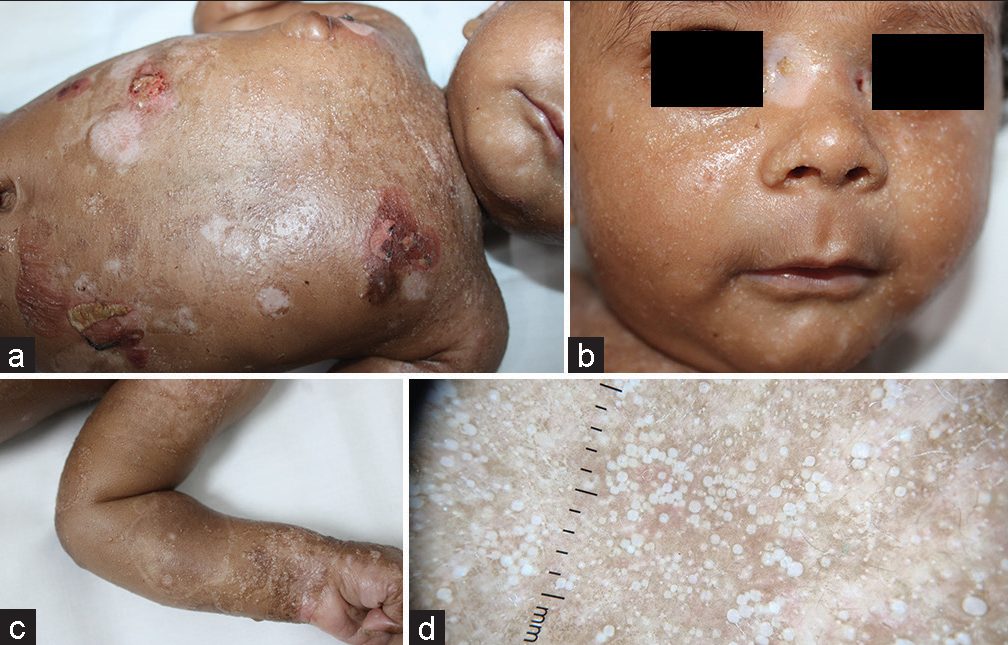
- (a) Multiple eruptive milia with milia en plaque noted over the chest, face, forearm. (b) Multiple eruptive milia noted over the face. (c) Multiple eruptive milia noted over the forearm. (d) Dermoscopy showing multiple monomorphic pearly white papules of milia (Dermlite DL 4, ×10).
Milia are small keratinous cysts of the epidermis that arise either de novo as primary milia or secondary to other underlying etiology as secondary milia. Primary milia arise from the sebaceous collar of vellus hairs in contrast to secondary milia which originates from the eccrine ducts. The genodermatoses associated with milia include genetic blistering diseases such as EB, Bazex syndrome, Brooke–Spiegler syndrome, and pachyonychia congenita. Among the EB, milia are usually seen in dystrophic subtypes associated with scarring and sometimes EB herpetiformis (Dowling Meara). As the split is present in sublamina densa in dystrophic EB, milia are more commonly seen. Hence, milia can be used as a clinical marker for differentiating between these different subtypes, as it is rarely observed in other phenotypes of hereditary EB.[1]
Declaration of patient consent
The authors certify that they have obtained all appropriate patient consent.
Conflicts of interest
There are no conflicts of interest.
Use of artificial intelligence (AI)-assisted technology for manuscript preparation
The authors confirm that there was no use of artificial intelligence (AI)-assisted technology for assisting in the writing or editing of the manuscript and no images were manipulated using AI.
Financial support and sponsorship
Nil.
References
- Milia: A review and classification. J Am Acad Dermatol. 2008;59:1050-63.
- [CrossRef] [PubMed] [Google Scholar]


