
Translate this page into:
Under the folds: Demystifying uncommon presentations of diaper dermatitis

*Corresponding author: Anubhab Bhattacharyya, Department of Dermatology, JIPMER, Puducherry, India. anubhabriorik@gmail.com
-
Received: ,
Accepted: ,
How to cite this article: Bhattacharyya A, Somasundaram A, Kotekar S. Under the folds: Demystifying uncommon presentations of diaper dermatitis. CosmoDerma. 2023;3:169. doi: 10.25259/CSDM_216_2023
Dear Sir,
Diaper rash represents a common clinical condition with varying presentations. While arriving at a diagnosis is not usually difficult, sometimes, the patient can present with findings not commonly encountered. We hereby describe two unusual variants/presentations of diaper rash, which were seen as two separate cases.
The first case was that of an 8-month-old male infant, who recently underwent surgery for anorectal malformation, presented with complaints of multiple raised solid lesions in the perianal region for a duration of two weeks following the surgical procedure. There is a history of diaper usage and fecal contamination. Cutaneous examination revealed the presence of ill-defined papules, plaques with surface erosions, and nodular lesions arranged symmetrically on either side of the anal orifice [Figure 1]. Tzanck smear, Gram stain, and potassium hydroxide mount were done from the lesions, and all were negative. Based on clinical findings, a diagnosis of perianal pseudoverrucous papules and nodules (PPPN) was considered.
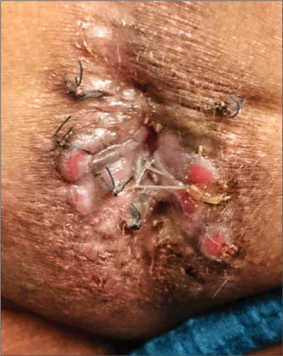
- Ill-defined papules, plaques with surface erosions, and nodular lesions arranged symmetrically on either side of the anal orifice.
The second case was that of a 15-day-old male neonate admitted to the neonatal intensive care unit for low birth weight, was noted to have developed multiple erosions over the buttocks by the caregivers, for which they sought dermatology consultation. We observed the use of a diaper in the child, which was visibly soiled with fecal matter. On examination, we found the presence of multiple discrete 1–2 mm well-defined superficial erosions with erythematous floor and irregular margins distributed symmetrically over the gluteal convexities [Figure 2]. Surrounding skin showed no erythema or pustules. Tzanck smear, Gram stain, and potassium hydroxide mount revealed nothing. Thus, based on presentation and examination, we arrived at a diagnosis of Jacquet’s erosive dermatitis.
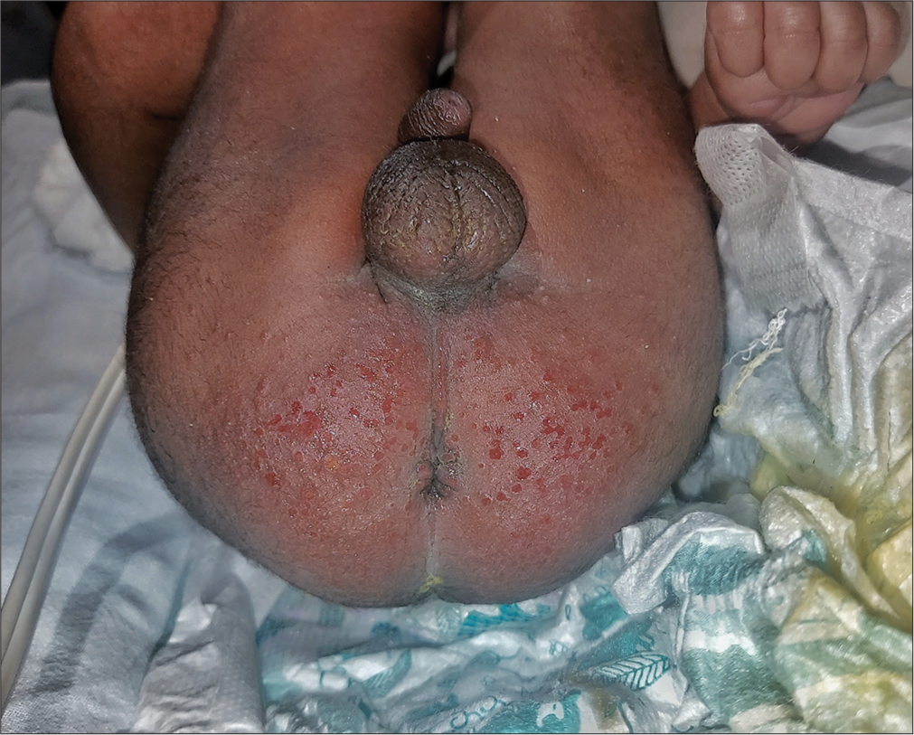
- Discrete 1–2-mm well-defined superficial erosion margins distributed symmetrically over the gluteal convexities.
Diaper dermatitis presentations include those of irritant contact dermatitis, allergic contact dermatitis, candidal and streptococcal intertrigo, and perianal streptococcal disease, while less common ones include granuloma gluteal infantum, Jacquet’s erosive dermatitis, and pseudoverrucous papules and nodules.[1] The reason for this specific pattern of involvement in this anatomic region of the body is multifactorial, which includes persistent fecal and urine contamination, friction, local humidity, temperature, and colonization by the gut, genitourinary or cutaneous flora, contact with chemical irritants. The previously hypothesized role of the presence of ammoniacal compounds in urine and feces is now refuted[1] – although the presence of several enzymes (such as fecal lipase and protease) and gut microbiome that is brought to the skin through fecal route play an important role in initiating or perpetuating the rash. In specific cases where there is some anatomical or surgical defect (for example, post-surgery in a patient with Hirschsprung’s disease) or persistent diarrhea or urinary leakage predisposing to continued exposure to and irritation of the skin by fecal matter/urine, the patient can develop such conditions as PPPN or Jacquet’s erosive dermatitis. Both entities have a common pathogenesis but their presentations are vastly different. While PPPN appears such as papules, nodules, and verrucous plaques on either side of genital/anal orifice with variable maceration,[2] Jacquet’s erosive dermatitis presents with shallow discrete ulcerations with islands of re-epithelialization over the buttocks.[3]
Treatment should be aimed at reducing the irritation and prevention of secondary infection by removal of precipitating factor. Restoration of the normal skin barrier is also central to the recovery process. Wherever applicable, the peristomal skin should be swabbed for microbiological culture as infections are treatable causes of complications.
These cases thus highlight the importance of knowing uncommon clinical presentations of diaper rash, especially in the presence of predisposing factors and where secondary causes have been ruled out.
Declaration of patient consent
The authors certify that they have obtained all appropriate patient consent.
Conflicts of interest
There are no conflicts of interest.
Use of artificial intelligence (AI)-assisted technology for manuscript preparation
The authors confirm that there was no use of artificial intelligence (AI)-assisted technology for assisting in the writing or editing of the manuscript and no images were manipulated using AI.
Financial support and sponsorship
Nil.
References
- Napkin dermatitis In: Hoeger PH, Kinsler V, Yan AC, Bodemer C, Larralde M, Luk D, eds. Harper’s textbook of pediatric dermatology. Vol 20. United States: John Wiley and Sons; 2019. p. :265-78.
- [Google Scholar]
- Perianal pseudoverrucous papules and nodules. Indian J Sex Transm Dis AIDS. 2013;34:44-6.
- [CrossRef] [PubMed] [Google Scholar]
- Jacquet's erosive dermatitis in an elderly individual: A rare manifestation of topical corticosteroid abuse. Indian Dermatol Online J. 2021;12:616-8.
- [CrossRef] [PubMed] [Google Scholar]


