
Translate this page into:
Cost-effective models for microsurgical training

*Corresponding author: Ravi Kumar Chittoria, Department of Plastic Surgery, Jawaharlal Institute of Postgraduate Medical Education and Research, Puducherry, India. drchittoria@yahoo.com
-
Received: ,
Accepted: ,
How to cite this article: Chittoria RK, Parthiban B, Prathyusha S. Cost-effective models for microsurgical training. CosmoDerma. 2023;3:168. doi: 10.25259/CSDM_208_2023
Abstract
In several surgical subspecialties, microsurgery training is essential. Many microsurgical training models were proposed for over a century. This includes live and non-viable tissue and organic and inorganic models. This investigation was done in a tertiary care plastic surgery department. The article reviews Scopus, PubMed, Google Scholar, and internet literature. We provide cost-effective strategies for teaching residents and juniors in microsurgery and maintaining surgeon skills. Super specialty residents worldwide need microsurgical training. Microsurgical training materials are scarce in certain locations. The foregoing readily available materials help residents practice continuously and consistently to improve their microsurgical confidence and skills.
Keywords
Microsurgery
Training
Models
Cost-effective
Umbilical cord

INTRODUCTION
Microsurgery is an important surgical field and microsurgery training continues to be fundamental in many surgical subspecialties. For over a century, various models were suggested for microsurgical training.[1,2] This includes organic and inorganic models and viable and non-viable tissue. We are presenting some cost-effective models that can be useful in training residents and juniors on microsurgery skills and also help surgeons maintain their skills.
It is a perspective article based on literature available in Scopus, PubMed, Google Scholar, and internet. Based on available literature, we have identified the following materials found to be useful in the microsurgical training among the residents.[3]
Surgical gloves
Bubble wrap
Human umbilical cord.
SURGICAL GLOVES
The surgical gloves are easily and readily available and accessible and can be used for microsurgery training such as suturing and tissue handling.[4] Gloves are stretched and applied over the plastic frame with a contrast background inside the gloves. The contrast background helps in differentiation of the suturing edges of the incised gloves. Plastic apron and color paper can be used as contrast background. It helps in improving our hand movement practice in microsurgery [Figure 1].
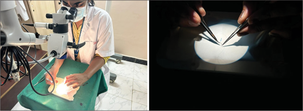
- Use of surgical gloves in microsurgery training.
Advantages
They are readily available, very cheap. The resident can undergo training at any place with gloves and magnification loupe.
Disadvantages
It does not have a human tissue-like feel. Tearing of gloves can occur.
BUBBLE WRAP
The bubble wrap is a cheap and easy-to-find training tool, with a thickness varying from 3/16 to 1-inch big bubbles. For laboratory training, the small-size bubbles are preferred. The bubble wrap comes with a thickness varying from 3/16 to 1-inch big bubbles. For laboratory training, the small-size bubbles are preferred. The very thin upper layer of plastic is very fragile and needs very careful handling during exercise [Figure 2]. In neurosurgery, the bubble wrap have the same fragility of the arachnoid or very small intracranial vessels, such as distal cortical vessels.[5]
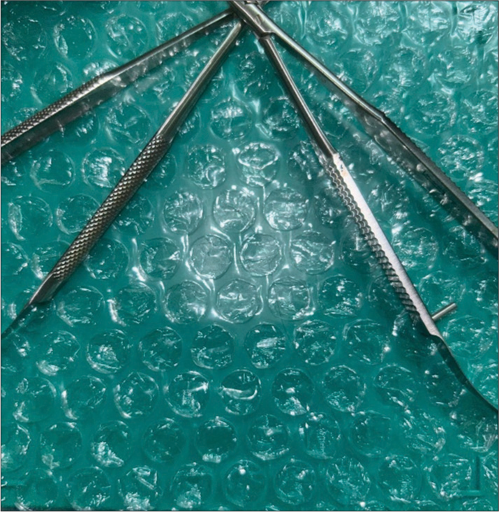
- Use of bubble wrap in microsurgery training.
Advantages
It is easily available, very cheap. It helps in learning tissue handling. It resembles similar human tissue.
Disadvantages
Difficulty in visualizing the suturing edges of the bubble wrap compared to gloves. It can be overcome by filling the bubble with contrast color fluid before practicing suturing.
UMBILICAL CORD
The umbilical cord is a lifeline between the fetus and the placenta and contains two arteries and one vein. The vessels lie in a supporting myxomatous tissue derived from the mesoderm, the “Wharton’s Jelly.” In the plastic surgery department at our hospital, we utilized the umbilical cord as a microsurgery-training model after acquiring a written consent from the mothers. The umbilical cord was brought from the obstetric unit and harvested, under strong restrictions regarding human immunodeficiency virus, hepatitis viruses, and other pathological infections to prevent infection transmission.[6] After harvesting clean with adequate saline to remove the blood clots. Methylene blue dye is used to fill the vessels of umbilical cord with cannula. The coloring of the umbilical cord helps in providing good contrast colors for easier practice [Figure 3].
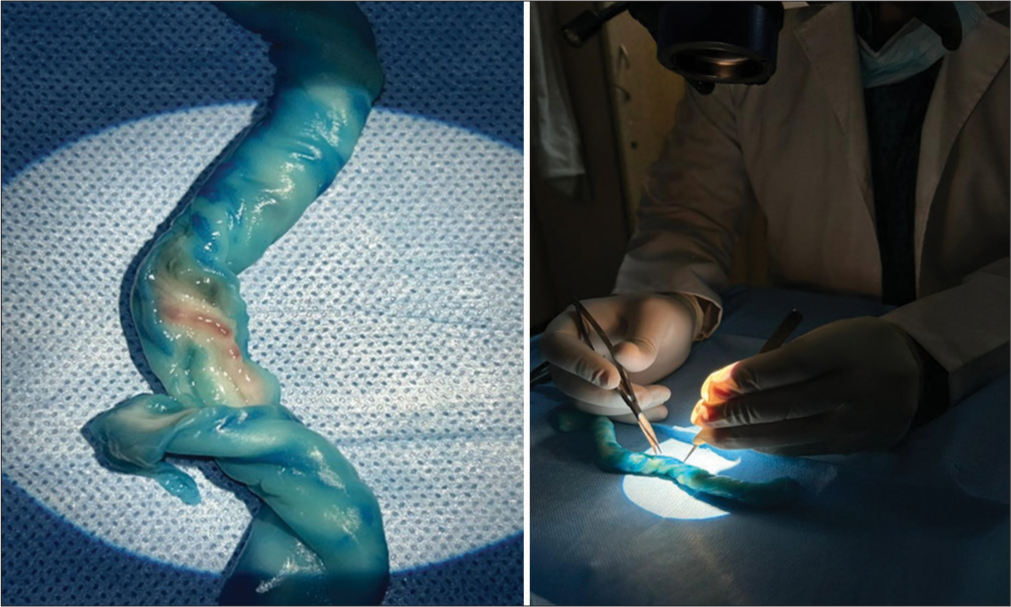
- Use of human umbilical cord in microsurgery training.
Advantages
It resembles the human tissue. It gives more expertise in practicing vessel dissection and followed by cutting the real vessel and suture it back.
Disadvantages
Not readily available. Practicing should be done in a proper place in hospital. It should be discarded safely. Serology screening is a must. Human umbilical cord was harvested after obtaining permission from institute, concerned department, and also from the delivering mother.
CONCLUSION
Microsurgical training is much needed in the super specialty residents all over the world. Microsurgical training materials are not widely available in all centers. The above materials which are easily available help the residents in continuous and consistent training for improving their confidence and skill set in microsurgical training.
Declaration of patient consent
The authors certify that they have obtained all appropriate patient consent.
Conflicts of interest
There are no conflicts of interest.
Use of artificial intelligence (AI)-assisted technology for manuscript preparation
The authors confirm that there was no use of artificial intelligence (AI)-assisted technology for assisting in the writing or editing of the manuscript and no images were manipulated using AI.
Financial support and sponsorship
Nil.
References
- Microsurgery training in plastic surgery. Plast Reconstr Surg Glob Open. 2020;8:e2898.
- [CrossRef] [PubMed] [Google Scholar]
- Validation of microsurgical models in microsurgery training and competence: A review. Microsurgery. 2007;27:494-9.
- [CrossRef] [PubMed] [Google Scholar]
- Microvascular surgical training models. J Plast Reconstr Aesthet Surg. 2011;64:e210-2.
- [CrossRef] [PubMed] [Google Scholar]
- The use of a surgical glove in microsurgical training: A new point of view. Microsurgery. 2010;30:505-6.
- [CrossRef] [PubMed] [Google Scholar]
- Use of bubble wrap for microsurgical training. J Reconstr Microsurg. 2013;29:635-6.
- [CrossRef] [PubMed] [Google Scholar]
- The human umbilical cord: A model for microsurgical training. J Hand Microsurg. 2014;6:111-2.
- [CrossRef] [PubMed] [Google Scholar]


