Generic selectors
Exact matches only
Search in title
Search in content
Post Type Selectors
Filter by Categories
Brief Report
Case Report
Case Series
Editorial
Focus
Images/Instrument in Dermatology/Dermatosurgery
Innovations
Letter to the Editor
Living Legends
Looking back in history
Original Article
Perspective
Resident Forum
Review Article
Spot the Diagnosis
Tropical Dermatology
Visual Treats in Dermatology
View/Download PDF

Translate this page into:

Visual Treats in Dermatology
2022
:2;
55
doi:
10.25259/CSDM_61_2022

Pseudomonas infection of nail: Green nail syndrome secondary to onychomycosis

Department of Dermatology, All India Institute of Medical Sciences Rishikesh, Rishikesh, Uttarakhand, India.
Author image

*Corresponding author: Department of Dermatology, All India Institute of Medical Sciences Rishikesh, Rishikesh, Uttarakhand, India. anmolbatra9@gmail.com

Licence
This is an open-access article distributed under the terms of the Creative Commons Attribution-Non Commercial-Share Alike 4.0 License, which allows others to remix, transform, and build upon the work non-commercially, as long as the author is credited and the new creations are licensed under the identical terms.

How to cite this article: Batra A, Hazarika N. Pseudomonas infection of nail: Green nail syndrome secondary to onychomycosis. CosmoDerma 2022;2:55.

A 21-year-old female presented with a 3-month history of slowly progressive greenish-black discoloration of right middle fingernail and left ring fingernail, with distolateral onycholysis of both middle fingernails and left ring fingernail with proximal nontender paronychia of both middle fingernails [Figure 1]. There was mild pus discharge from undersurface of left middle fingernail as well.
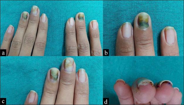
Proximal greenish-black discoloration, distolateral onycholysis, and proximal paronychia seen over (a) both middle fingernails and left ring fingernail. (b) Close-up view of right middle fingernail. (c) Close-up view of left middle and ring fingernail. (d) Mild pus discharge from undersurface of left middle fingernail.
Figure 1:
Proximal greenish-black discoloration, distolateral onycholysis, and proximal paronychia seen over (a) both middle fingernails and left ring fingernail. (b) Close-up view of right middle fingernail. (c) Close-up view of left middle and ring fingernail. (d) Mild pus discharge from undersurface of left middle fingernail.

Direct mycologic examination of nail scrapings was positive for fungal elements and bacteriologic examination of nail scrapings revealed Pseudomonas aeruginosa infection. She was prescribed itraconazole 200 mg/day 7 days per month for 2 months for fungal infection and oral ciprofloxacin 500 mg/day for 3 weeks for pseudomonas coverage. There was significant improvement on follow-up.

P. aeruginosa is the most common pathogen causing bacterial nail infections.[1] Treatment options include topical gentamicin for 1-3 months or oral ciprofloxacin for 2-3 weeks.

Declaration of patient consent

Patient’s consent not required as patient’s identity is not disclosed or compromised.

Financial support and sponsorship

Nil.

Conflicts of interest

There are no conflicts of interest.

References

  1. , , . Infections of finger and toe nails due to fungi and bacteria. Hautarzt. 2014;65:337-48.
    [CrossRef] [PubMed] [Google Scholar]

Fulltext Views
5,330

PDF downloads
1,092
View/Download PDF
Download Citations
BibTeX
RIS
Show Sections