
Translate this page into:
Geometric nail pitting in alopecia areata

*Corresponding author: Sharang Gupta, Department of Dermatology, Government Medical College, Patiala, Punjab, India. drsharanggupta97@gmail.com
-
Received: ,
Accepted: ,
How to cite this article: Gupta S, Chopra D. Geometric nail pitting in alopecia areata. CosmoDerma 2022;2:53.
A 17-year-old boy presented with the chief complaints of patchy hair loss in the scalp for 3 years. On examination, multiple, smooth, well-defined patches of alopecia were appreciable on the scalp. Trichoscopy of the patches revealed multiple black dots, yellow dots, exclamation mark hair, and Pohl-Pinkus constrictions. He was diagnosed as a case of alopecia areata. On examination of the nails, multiple small superficial pits were seen in the nail plate which were regularly distributed in a geometric pattern along the longitudinal and transverse lines. The pattern was better visualized with the help of dermoscopic examination at ×10 magnification (non-polarized, contact mode) [Figure 1].
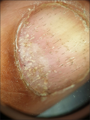
- Onychoscopic examination showing regular superficial pits in the nail plate (Hiene Delta 20T dermatoscope, ×10, non-polarized, contact mode).
Pits are defined as superficial depressions in the nail plate. They are formed due to sloughing off of clusters of parakeratotic cells in the uppermost layers of the nail plate when exposed to the external environment. Nail pitting is associated with many dermatological disorders, among which alopecia areata forms a common cause. The pits in alopecia areata are superficial and present in a geometric pattern along the longitudinal and transverse lines.[1] Onychoscopy serves as an easy-to-use and noninvasive tool to identify subtle changes in the nail unit.
Declaration of patient consent
Patient’s consent not required as patient’s identity is not disclosed or compromised.
Financial support and sponsorship
Nil.
Conflict of interest
There are no conflicts of interest.
References
- Nail pitting and onycholysis. Indian J Dermatol Venereol Leprol. 2009;75:631-3.
- [CrossRef] [PubMed] [Google Scholar]


