
Translate this page into:
Giant dermatofibroma on the ankle

*Corresponding author: Pradeep S. Nair, Department of Dermatology and Venereology, Government T D Medical College Alappuzha, Kerala, India. dvmchtvm@yahoo.co.in
-
Received: ,
Accepted: ,
How to cite this article: Nair PS, Nandana TV. Giant dermatofibroma on the ankle. CosmoDerma. 2023;3:188. doi: 10.25259/CSDM_254_2023.
A 16-year-old female presented with a hyperpigmented nodule on the left ankle of one-year duration. The lesion first started as a small asymptomatic brownish-black papule, which later progressed to form the present size with increase in pigmentation. There was no history of any nevus at the site preceding the present lesion. On examination, there was a well-defined hyperpigmented nodule of 3 cm in diameter on the upper left ankle with the surface showing tiny pits [Figure 1]. Dimple sign was negative. Dermoscopy showed a central white area with a few blood vessels, which were nonspecific. An excision skin biopsy showed minimal epidermal changes while the dermis showed a grenz zone with the dermis packed with spindle-shaped histiocytes arranged in whorls and circles in a storiform pattern [Figure 2]. High-power (× 400) magnification showed multinucleate giant cells with blood vessels [Figure 3], all diagnostic of dermatofibroma. Dermatofibromas are benign tumors of the fibrous tissue and may mimic melanoma clinically. Cellular and aneurysmal types are the other histological variants.[1] Dermascopy is non-specific but may show central scar, shiny white lines, and atypical vessels. Rarely, malignant transformation to dermatofibrosarcoma protuberans may occur, which stains positive for CD34. Surgical excision is the treatment of choice.[1]
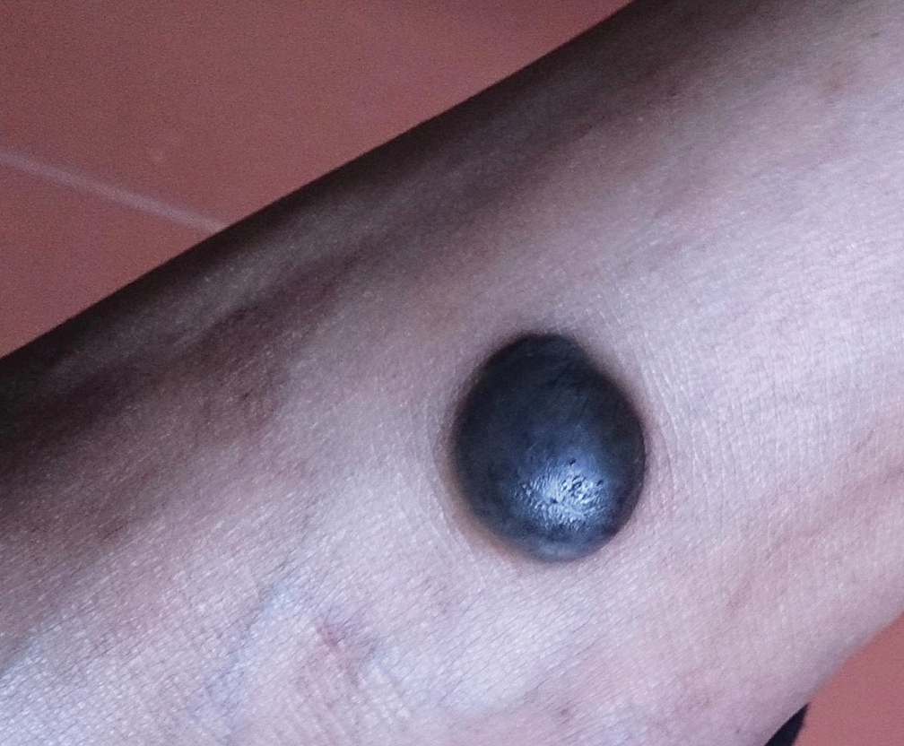
- Well-defined hyperpigmented nodule on left ankle.
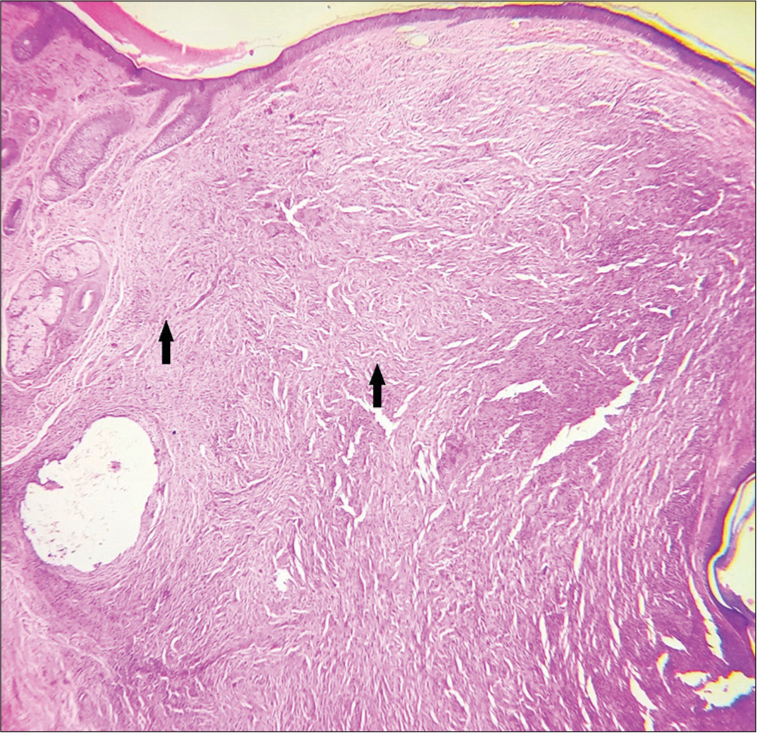
- Skin biopsy showing grenz zone, spindle-shaped histiocytes (arrows) arranged in storiform pattern, H and E × 100.
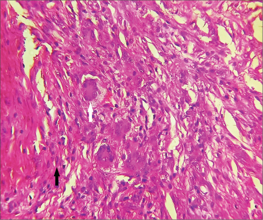
- Skin biopsy (High power) showing giant cells (white arrow), blood vessels, and spindle-shaped histiocytes (black arrow), H and E × 400.
Ethical Approval
Institutional Review Board approval is not required.
Declaration of patient consent
The authors certify that they have obtained all appropriate patient consent.
Conflicts of interest
There are no conflicts of interest.
Use of artificial intelligence (AI)-assisted technology for manuscript preparation
The authors confirm that there was no use of artificial intelligence (AI)-assisted technology for assisting in the writing or editing of the manuscript and no images were manipulated using AI.
Financial support and sponsorship
Nil.
References
- Cellular dermatofibroma: Clinicopathological review of 218 cases of cellular dermatofibromas to determine clinical recurrence rate. Dermatol Surg. 2019;45:1359-64.
- [CrossRef] [PubMed] [Google Scholar]


