
Translate this page into:
Giant congenital melanocytic nevus

*Corresponding author: Suruthi Purushothaman, Department of Dermatology, Jawaharlal Institute of Postgraduate Medical Education and Research, Puducherry, India. suruthidvlpub@gmail.com
-
Received: ,
Accepted: ,
How to cite this article: Purushothaman S, Dharanisankar S. Giant congenital melanocytic nevus. CosmoDerma 2023;3:43.
A 2-year-old male boy born out of a consanguineous marriage by a normal vaginal delivery at term, with an uneventful antenatal history, presented to the dermatology outpatient department with an extensive black-pigmented patch over trunk since birth. There was no family history of any similar lesion. Cutaneous examination revealed an extensive hyperpigmented patch covering 50% of the back and extending to the front of the abdomen [Figures 1 and 2]. A single cerebriform nodule of size measuring 3×3 cm was present on the pigmented patch over the left lower back. Multiple hyperpigmented satellite lesions of size 4–5 cm were also present all over the body, extremities, and left ear lobule. Tufts of coarse and lustreless hair were scattered all over the lesion. There were no other associated congenital anomalies. Ultrasound of the abdomen and radiological examination of the spine were normal. Histopathological findings were consistent with those of congenital melanocytic naevi. No evidence of a malignant transformation was seen and yearly follow-up was advised.
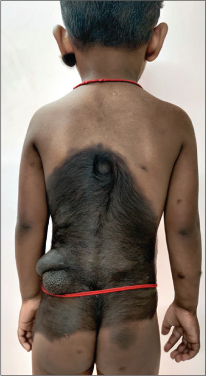
- Extensive pigmented patch covering 50% of the skin surface area over the back. Note the cerebriform nodule pigmented patch over the left lower back.
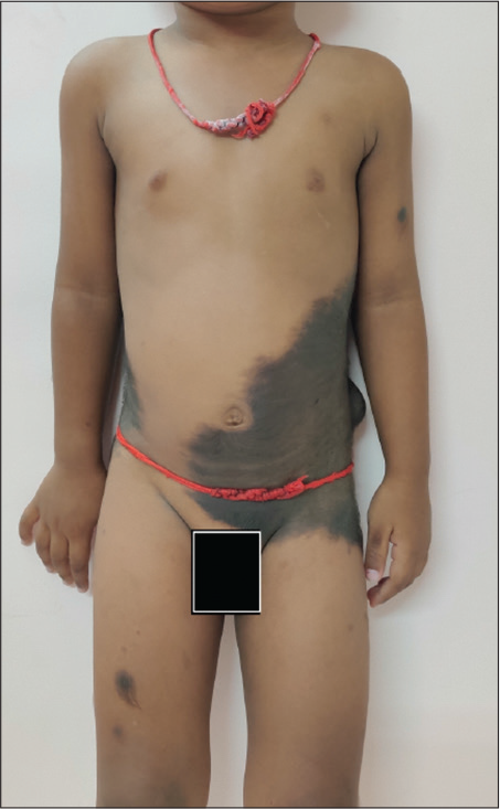
- Pigmented patch extending to the front of the abdomen.
Declaration of patient consent
The authors certify that they had obtained all appropriate consent from patients legally accepted guardian.
Conflicts of interest
There are no conflicts of interest.
Financial support and sponsorship
Nil.


