
Translate this page into:
Frog-spawn appearance: Diagnostic clue to lymphangioma circumscriptum


*Corresponding author: Anubhab Bhattacharyya, Department of Dermatology, JIPMER, Puducherry, India. anubhabriorik@gmail.com
-
Received: ,
Accepted: ,
How to cite this article: Bhattacharyya A, Sahadevan G. Frog-spawn appearance: Diagnostic clue to lymphangioma circumscriptum. CosmoDerma 2023;3:137.
An 11-year-old child came with multiple bubble-like lesions over the right upper thigh (near groin) for 2 years, which are asymptomatic for the most part except for occasional weeping of fluid from the site. During the current presentation, the child had no pain, weeping of fluid or symptoms suggestive of superadded infection. Parents gave no history of preceding trauma or any surgical corrective procedure undertaken. Cutaneous examination revealed the presence of three separate clusters of clear fluid-filled vesicles over the anterior aspect of the thigh [Figures 1a and b]. One vesicle around 3 mm in diameter showed the presence of blood-tinged fluid. A dermoscopic examination was performed, and clear and blood-tinged fluid-filled vesicles apparent as “lacunae” were noted [Figure 1c]. The characteristic morphology and dermoscopic appearance likened to a “frog-spawn” aided in making the diagnosis of microcystic lymphatic malformation (LM) or lymphangioma circumscriptum.
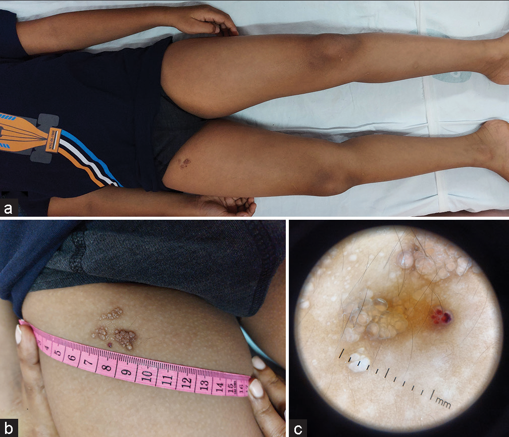
- (a and b) showing the location of the fluid-filled vesicles over the right anterior thigh, without any background of ill-defined swelling and (c) shows the dermoscopic appearance of fluid-filled lacunae with a single vesicle showing blood-tinged fluid content.
LM is a benign accumulation of lymph fluid in aberrant lymphatic channels and depending on the size of the swelling, it can be a microcystic, macrocystic, or mixed type. The onset can be at birth, or within the first 2–3 years of life, though cases with a later onset have also been reported. Microcystic LM are localized usually over skin or mucosa and can undergo secondary changes such as bleeding or verrucous changes.[1] Underlying lymphatic vessels occupy the dermal papilla and push upward against the epidermis, leading to a saccular appearance of the vesicles, which are described as “frog-spawn appearance.” Clinical identification of such lesions can help to avoid a biopsy and also to decide on the treatment modality. While surgical excision is the gold standard, for most cases outpatient care-based procedures such as radiofrequency ablation, ablative LASERs, cryotherapy, or sclerotherapy are employed. A local Doppler-aided ultrasound imaging helps to delineate the underlying extent of the malformation. Radiotherapy is usually not preferred because of the risk of malignant transformation. Treatment may need to be repeated if recurrence occurs.[1]
Declaration of patient consent
The authors certify that they have obtained all appropriate patient consent.
Conflicts of interest
There are no conflicts of interest.
Use of artificial intelligence (AI)-assisted technology for manuscript preparation
The authors confirm that there was no use of artificial intelligence (AI)-assisted technology for assisting in the writing or editing of the manuscript and no images were manipulated using AI.
Financial support and sponsorship
Nil.
References
- Cutaneous lymphangioma circumscriptum: Frog spawn on the skin. Int J Dermatol. 2009;48:1290-5.
- [CrossRef] [PubMed] [Google Scholar]


