
Translate this page into:
Dahlia flower appearance on dermoscopy

*Corresponding author: Spandana Devarahalli Krishnamurthy, Department of Dermatology, Bangalore Medical College, Bengaluru, Karnataka, India. spandanasinchu@gmail.com
-
Received: ,
Accepted: ,
How to cite this article: Krishnamurthy SD, Hanjagi P. Dahlia flower appearance on dermoscopy. CosmoDerma 2023;3:102.
A 24-year-old male presented with an asymptomatic raised skin lesion over the tip of the nose and philtrum of the upper lip from the past 6 months. No history of similar lesions elsewhere in the body. On dermatological examination, two filiform papules of size 2 mm and 5 mm in diameter, pedicled base, and rough surface were noted over the tip of the nose and philtrum of the upper lip, respectively [Figure 1]. The polarized dermoscopy of the lesion revealed a filiform feature of the wart, which was centered by a linear vessel that followed the direction of the filiform extension to the top [Figure 2]. The image was compared to dahlia flower-like pattern with a tapered finger pattern, a keratotic white tip, and pigmented zones [Figure 3].[1]
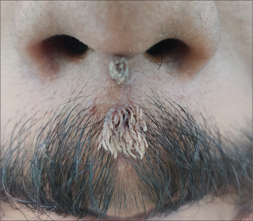
- Filiform papules of size 2 mm and 5 mm noted over tip of the nose and philtrum of the upper lip, respectively.
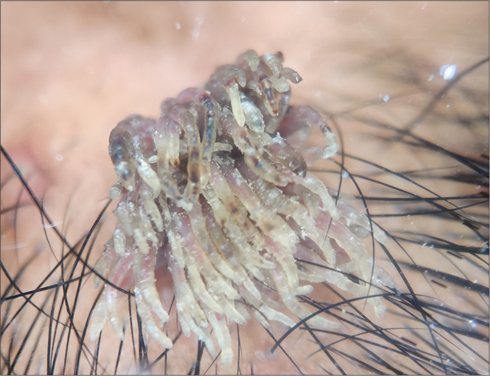
- Folarized dermoscopy of the lesion revealed a filiform feature similar to Dahlia flower with a tapered finger pattern, a keratotic white tip, and pigmented zones.
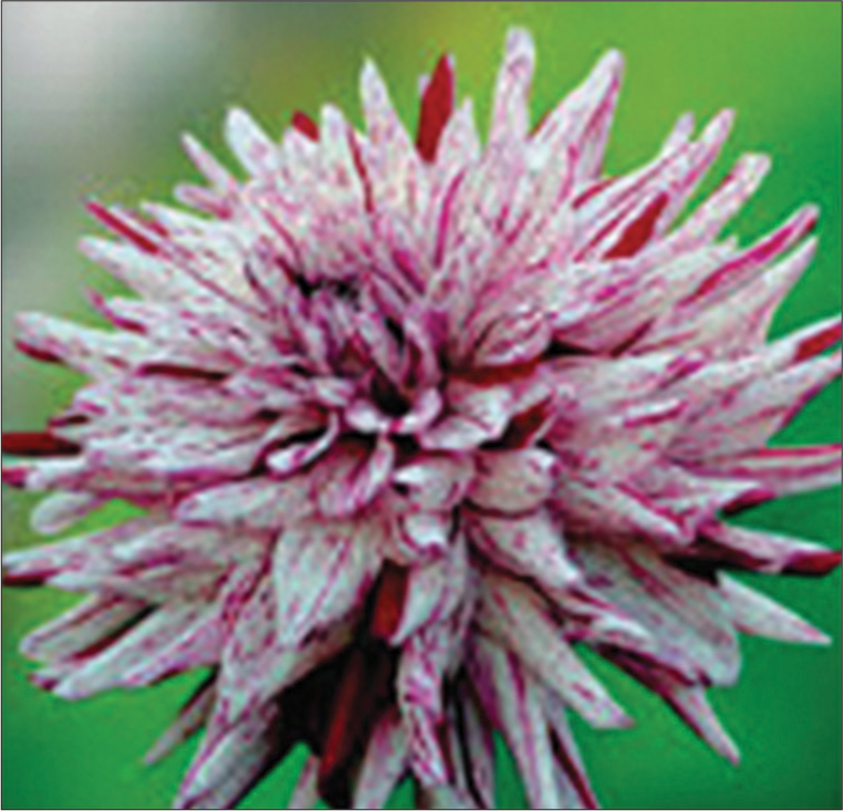
- Dahlia flower.
Warts are common benign epidermal growths resulting from different human papillomavirus strains. They are often categorized according to their morphology, histology, and anatomical location. A morphological type of wart termed verruca filiformis, 4% of the warts are filiform warts[2] which can occasionally have a small base and longer, finger-like extensions on the face and limbs. The diagnosis of viral warts is typically made clinically; however, dermoscopy can be helpful if the diagnosis is unclear or if it has to be distinguished from other conditions, such as seborrheic keratoses. It reveals filiform extensions with dilated blood vessels in the center that follows the path of these extensions at the summits, giving the impression of a hairpin. Dermoscopy may enhance the precision of detecting various clinically non-classical cutaneous wart types and aid in separating them from other overlapping skin diseases.
Declaration of patient consent
Patient’s consent not required as patients identity is not disclosed or compromised.
Conflicts of interest
There are no conflicts of interest.
Financial support and sponsorship
Nil.
References
- A dahlia like pattern: A new dermoscopic sign of filiform wart. Our Dermatol Online. 2021;12:e47.
- [CrossRef] [Google Scholar]
- The rare dermoscopic image of a filiform wart of lip in child. Ann Clin Case Stud. 2020;2:1022.
- [Google Scholar]


