
Translate this page into:
Carcinoma erysipeloides

*Corresponding author: Sharang Gupta, Department of Dermatology, Government Medical College, Patiala, Punjab, India. drsharanggupta97@gmail.com
-
Received: ,
Accepted: ,
How to cite this article: Gupta S, Chopra D. Carcinoma erysipeloides. CosmoDerma 2022;2:56.
A 56-year-old female presented with the chief complaint of a raised red lesion over the left breast for 6 months. The patient had undergone modified radical mastectomy of the left breast 2 years back owing to a diagnosis of invasive lobular carcinoma breast after which the patient did not receive any other form of treatment. On examination, a well-defined, erythematous and indurated plaque was present on the left breast [Figure 1]. The local temperature of the lesion was not raised. The scar of the previous surgery was appreciable on the left breast. No swelling of the left limb was noted. Examination of the right breast did not reveal any abnormal findings. The rest of the cutaneous and systemic examination was within normal limits. Histopathological examination revealed deposition of tightly packed malignant cells within the superficial and deep lymphatics. The patient was labeled as a case of carcinoma erysipeloides. The patient has been planned for Etoposide and Daunorubicin chemotherapy.
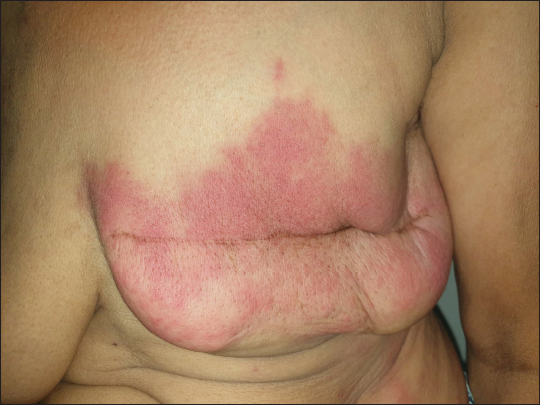
- A well-demarcated, erythematous, and indurated plaque present over the left breast.
Carcinoma erysipeloides is a form of cutaneous metastasis from underlying internal malignancy, most commonly breast carcinoma. Cutaneous metastasis can present in innumerable forms and can mimic a variety of conditions. Carcinoma erysipeloides can mimic erysipelas, cellulitis, and mastitis on the first look. Cases like these underscore the need for careful history taking, and clinical and histopathological examination, more so in patients with a history or risk factors of internal malignancies.[1]
Declaration of patient consent
Patient’s consent not required as patients identity is not disclosed or compromised.
Financial support and sponsorship
Nil.
Conflict of interest
There are no conflicts of interest.
References
- Post-mastectomy breast rash. Carcinoma erysipeloides. Int J Dermatol. 2010;49:855-7.
- [CrossRef] [PubMed] [Google Scholar]


