Translate this page into:
Alopecia totalis in an infant

*Corresponding author: Ilavendiran Sekar, Department of Dermatology, Velammal Medical College, Madurai, Tamil Nadu, India. s.ila3667@gmail.com
-
Received: ,
Accepted: ,
How to cite this article: Sivakumar A. Sekar I. Alopecia totalis in an infant. CosmoDerma 2023;3:41.
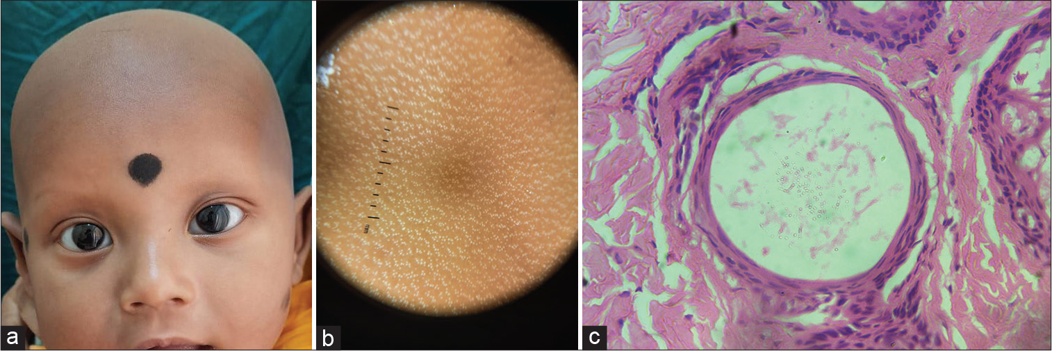
A 4-month-old male child was brought with complete loss of scalp and eyebrow hair since birth. The child was born of third-degree consanguineous marriage, but no similar complaints in the family were present. No other systemic comorbidities or any drug intake preceding the hair loss was present. Antenatal history was uneventful, and the child was born at term without any complications. On examination, complete alopecia of the scalp and eyebrows, and eyelashes were noted [Figure 1a]. No papules were seen, and nails and mucosa were within normal limits. A dermoscopy of the scalp revealed the presence of evenly distributed clusters of follicles throughout [Figure 1b]. A scalp biopsy sent for histology that revealed the absence of mature telogen follicles and the presence of multiple keratin cysts [Figure 1c], there was no inflammatory infiltrate in the dermis. Serum vitamin D levels and parathormone levels were normal in the patient. Hence, a diagnosis of atrichia with papular lesions was made.

- (a) Complete alopecia involving the scalp, eyebrows, and eyelashes, (b) dermoscopy (DL4, ×10, Polarized) showing cluster of stars appearance of hair follicular openings, and (c) high-power view (×400) of H and E specimen of scalp histology showing the presence of keratinous cysts of hair follicles.
Atrichia with papular lesions is an autosomal recessive disorder due to a mutation in the Hairless (HR) gene. HR is a hair cycle regulator; thereby, mutation causes defective catagen arrest, thereby unable to re-enter the anagen phase. This leads to progressive keratin accumulation and cyst formation histologically before becoming evident on clinical examination as papular lesions. Differentials to be considered should be alopecia universalis congenita and vitamin D dependent rickets type 2. Biochemical parameters such as serum calcium, alkaline phosphatase, parathormone, and calcium levels can be assessed along with bone radiography to rule out vitamin D dependent rickets. Gene studies for HR mutation can confirm the diagnosis which could not be done in our patient due to affordability issues. A biopsy may be useful in the early infancy to distinguish between these clinical entities when the papules are not yet fully developed.[1]
Declaration of patient consent
The authors certify that they have obtained all appropriate patient consent.
Conflicts of interest
There are no conflicts of interest.
Financial support and sponsorship
Nil.
References
- Atrichia with papular lesions: Importance of histology at an early disease stage. Skin Appendage Disord. 2018;4:129-30.
- [CrossRef] [PubMed] [Google Scholar]


