
Translate this page into:
Tinea localizing to vitiligo patch - An interesting observation

*Corresponding author: Arun Somasundaram, Department of Dermatology and STD, JIPMER, Puducherry, India. arunsomasundaram25@gmail.com
-
Received: ,
Accepted: ,
How to cite this article: Ramesh N, Somasundaram A, Sivakumar A. Tinea localizing to vitiligo patch - An interesting observation. CosmoDerma 2023;3:159.
Dear Sir,
Vitiligo is an autoimmune disorder. Tinea corporis is a superficial fungal infection. There are very few case reports of tinea being localized exclusively to a vitiligo patch. Here, we describe a case of tinea localizing a vitiligo patch.
A 77-year-old male presented with complaints of an itchy scaly lesion over his back for the past 1-month duration. He was a known case of vitiligo vulgaris for the past 9 years and was currently not on any treatment for the same. He had no other known comorbidities, including diabetes, and was not on any medication. There were no similar cutaneous complaints in his family members. Cutaneous examination revealed multiple depigmented macules distributed diffusely over his face, trunk, abdomen, back, and upper and lower limbs. The depigmented macule on his upper back was erythematous and scaly. It had an erythematous raised active border studded with pustules [Figure 1a], suggestive of dermatophytosis. There was no clinical evidence of dermatophyte infection over other vitiligo lesions or other parts of the body. Scrapings from the margin of the plaque as well as the pustules revealed fungal elements on potassium hydroxide 10% mount [Figure 1b]. A clinical diagnosis of tinea encircling vitiligo was offered. The patient was started on itraconazole and topical luliconazole lotion and was advised to follow-up after a month.
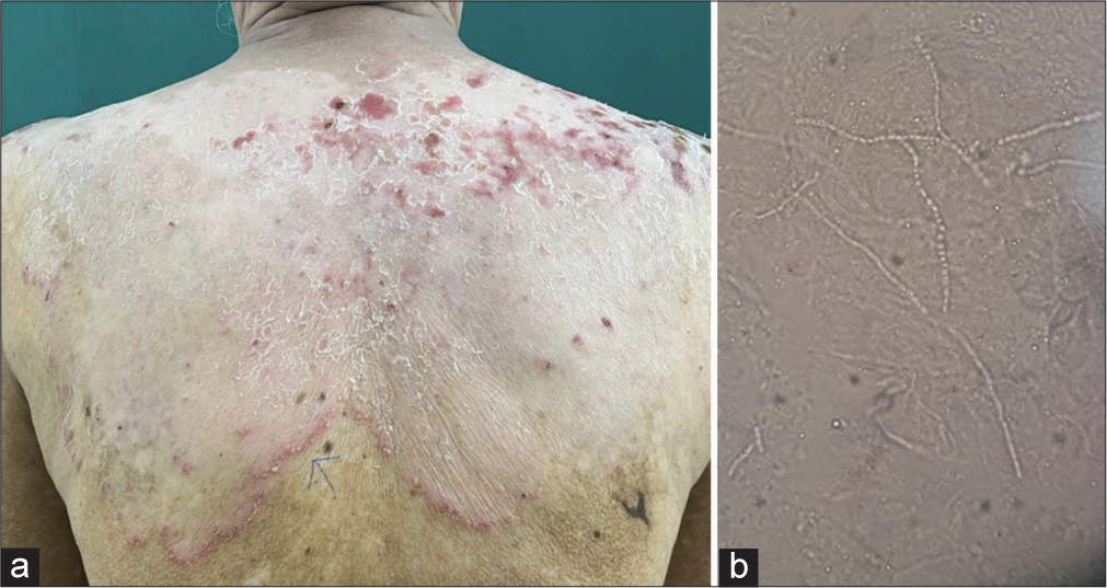
- (a) Depigmented macule over the upper back with erythematous raised active border studded with pustules. (b) Fungal elements on potassium hydroxide 10% mount.
Dermatophyte infection bordering or localizing only to a vitiligo patch is underrecognized with very few case reports in the literature. Various mechanisms have been proposed.[1-3] There is evidence that Langerhans cells are decreased in number and functionally impaired in vitiligo patches.[4] The occurrence of dermatophytosis in vitiligo patches might be due to the local immune defects either directly to reduced or absent melanocytes or indirectly to the reduced number and function of Langerhans cells in a vitiligo patch. These serve as antigen-presenting cells for dermatophytes and are thereby crucial in inducing T-lymphocyte response and eliminating dermatophytic infection.[2] Another proposed mechanism is that there may be alteration in the antigenicity of dermatophyte-infected keratinocytes at the periphery but not within vitiligo lesions, which is recognized by specific T-cells responsible for peripheral localization of dermatophyte infection in the vitiligo lesion.[1] One other theory is that of the actinic theory in which ultraviolet radiation induces recruitment of inflammatory cells such as neutrophils and monocytes into the vitiliginous area and not the normally pigmented skin, leading to the death of dermatophytic conidia within the lesion and residual infection at the border of the vitiligo patch.[1] Dermatophytosis occurring over a vitiligo patch may also be an expression of Wolf ’s isotopic response.[2,4,5] However, the involvement of only a single lesion of vitiligo is puzzling, as the fungal infection occurred on a vitiligo patch.
In conclusion, several theories have been put forth to explain the phenomenon of dermatophyte infection encircling and localizing to vitiligo. This condition rather than being rare is more underrecognized. This case highlights an interesting observation in our day-to-day practice and the need to be more aware of colocalizing dermatoses that exist.
Declaration of patient consent
Patient’s consent was not required as patient’s identity was not disclosed or compromised.
Conflicts of interest
There are no conflicts of interest.
Use of artificial intelligence (AI)-assisted technology for manuscript preparation
The authors confirm that there was no use of artificial intelligence (AI)-assisted technology for assisting in the writing or editing of the manuscript and no images were manipulated using AI.
Financial support and sponsorship
Nil.
References
- Dermatophyte infection encircling vitiligo. Indian Dermatol Online J. 2015;6:S60-3.
- [CrossRef] [PubMed] [Google Scholar]
- Vitiligo delimiting dermatophyte infection. Indian J Dermatol. 2015;60:91-3.
- [CrossRef] [PubMed] [Google Scholar]
- Could it be wolf isotopic response?: Occurrence of dermatophytoses on vitiliginous skin-case series of five cases from a tertiary care centre in India. Indian Dermatol Online J. 2022;13:240-3.
- [CrossRef] [PubMed] [Google Scholar]
- Evaluation of the Langerhans cells role in vitiligo and its relationship to NBUVB. J Cosmet Dermatol. 2021;20:3642-8.
- [CrossRef] [PubMed] [Google Scholar]


