
Translate this page into:
Segmental nevus depigmentosus

-
Received: ,
Accepted: ,
How to cite this article: Devi MS, Laishram R. Segmental nevus depigmentosus. CosmoDerma 2022;2:31.
A 6-year-old boy presented with whitish colored lesion over the right side of the face at 2 years of age. The lesion was gradually increasing in size with the child’s growth and there was no loss of sensation. On cutaneous examination, a solitary well-defined off-white macule with an irregular border extends from the right forehead to the malar area and nose with sharp demarcation in the midline. Smaller macules were present around the margin giving a ‘splashed paint’ appearance along with poliosis of the eyebrow [Figure 1]. Diascopy was negative and Wood’s lamp examination revealed an off-white color. Dermoscopy of the lesion shows irregular whitish areas with few areas of reticular pigment network. The irregular extensions are seen as pseudopods at the periphery with perifollicular pigmentation and there is the absence of perifollicular hyperpigmentation [Figure 2]. Based on the criteria given by Coup, a diagnosis of nevus depigmentosus was made. Nevus depigmentosus is a pigmentary disorder that presents with a nonprogressive sharply demarcated hypopigmented macule usually visible at birth or early childhood.[1]
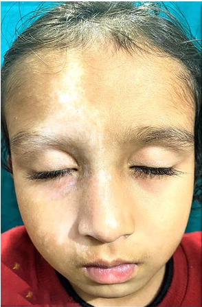
- Solitary well-defined off-white macule with irregular border and multiple similar small macules at the margin (‘splashed paint appearance’), extending from the right forehead to the right malar area and nose with sharp demarcation in midline.
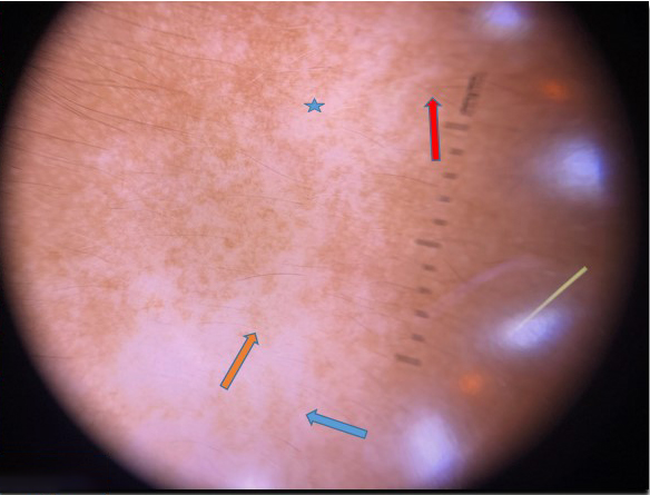
- Dermoscopy of the lesion shows irregular whitish areas (star), reticular pigment network (blue arrow), pseudopods at the periphery (red arrow), and perifollicular pigmentation (orange arrow) (3Gen DermLite IV Polarized Dermoscope).
Declaration of patient consent
The authors certify that they have obtained all appropriate patient consent.
Financial support and sponsorship
Nil.
Conflicts of interest
There are no conflicts of interest.
References
- Clinical and histopathological characteristics of nevus depigmentosus. J Am Acad Dermatol. 2006;55:423-8.
- [CrossRef] [PubMed] [Google Scholar]


