
Translate this page into:
Segmental hairy nevus

*Corresponding author: Rachita S. Dhurat, Department of Dermatology, Lokmanya Tilak Municipal Medical College and General Hospital, Mumbai, Maharashtra, India. rachitadhurat@yahoo.co.in
-
Received: ,
Accepted: ,
How to cite this article: Khandare M, Kowe PA, Dhurat RS. Segmental hairy nevus. CosmoDerma. 2024;4:37. doi: 10.25259/CSDM_25_2024
A 9-year-old girl presented with a complaint of a dark-colored lesion over her forehead since birth associated with thick hair. There was no history of seizures, and general and systemic examination was unremarkable. Cutaneous examination revealed a hyperpigmented mamillated plaque of size 10 cm × 8 cm associated with overlying thick coarse terminal hair present over the left forehead extending between the left eyebrow down to the left frontoparietal region without crossing the midline [Figures 1 and 2]. There was no evidence of ulceration, bleeding or nodularity. Based on history and clinical findings, a final diagnosis of segmental hairy nevus (congenital melanocytic nevus) was made. Parents were counseled about the prognosis of the nevus, and she was referred to a plastic surgeon for surgical excision.
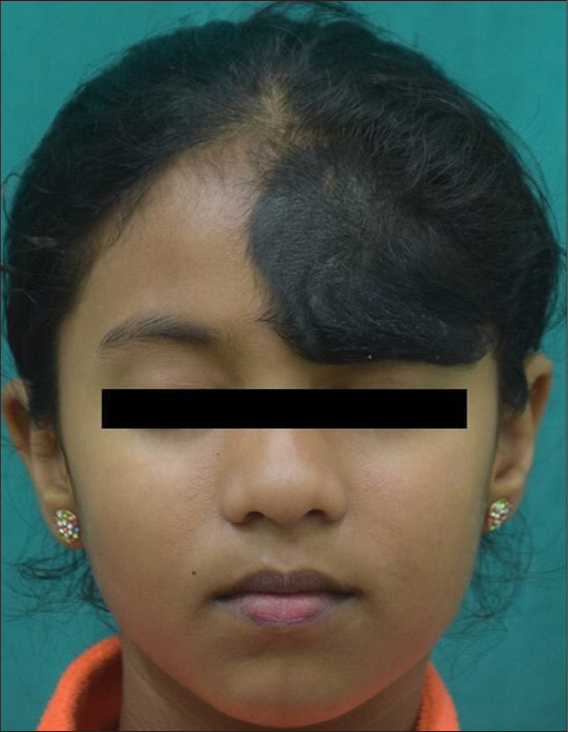
- Nevus with overlying hypertrichosis over the left side of the forehead.
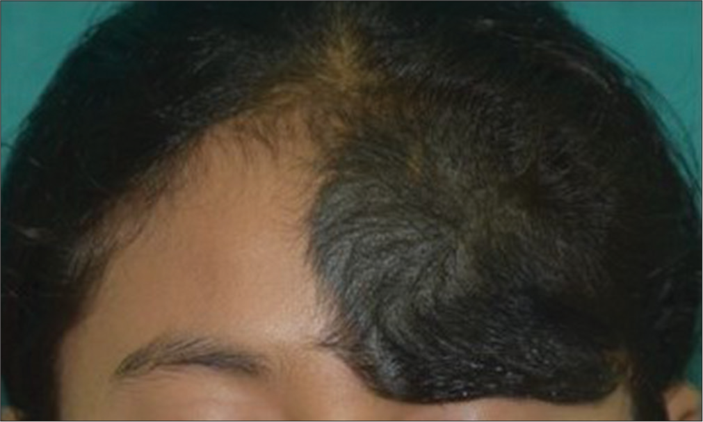
- Closer view of the nevus.
Ethical approval
The Institutional Review Board approval is not required.
Declaration of patient consent
The authors certify that they have obtained all appropriate patient consent.
Conflicts of interest
There are no conflicts of interest.
Use of artificial intelligence (AI)-assisted technology for manuscript preparation
The authors confirm that there was no use of artificial intelligence (AI)-assisted technology for assisting in the writing or editing of the manuscript and no images were manipulated using AI.
Financial support and sponsorship
Nil.


