
Translate this page into:
Scrotal calcinosis


*Corresponding author: Anmol Batra, Department of Dermatology, AIIMS Rishikesh, Rishikesh, Uttarakhand, India. anmolbatra9@gmail.com
-
Received: ,
Accepted: ,
How to cite this article: Batra A, Hazarika N. Scrotal calcinosis. CosmoDerma 2022;2:49.
A 24-year-old man presented with a 5-year history of numerous asymptomatic nodules of the scrotal skin which had been slowly progressing in number and size. Physical examination revealed multiple discrete and confluent, white-yellowish subcutaneous nodules measuring 0.2-2.5 cm in diameter attached to the scrotal skin [Figure 1]. The nodules were nontender and firm on palpation. Histopathological examination revealed calcium deposition within the dermis. Laboratory parameters did reveal any calcium/phosphorus imbalance. Based on the above characteristic description, a diagnosis of scrotal calcinosis was made.
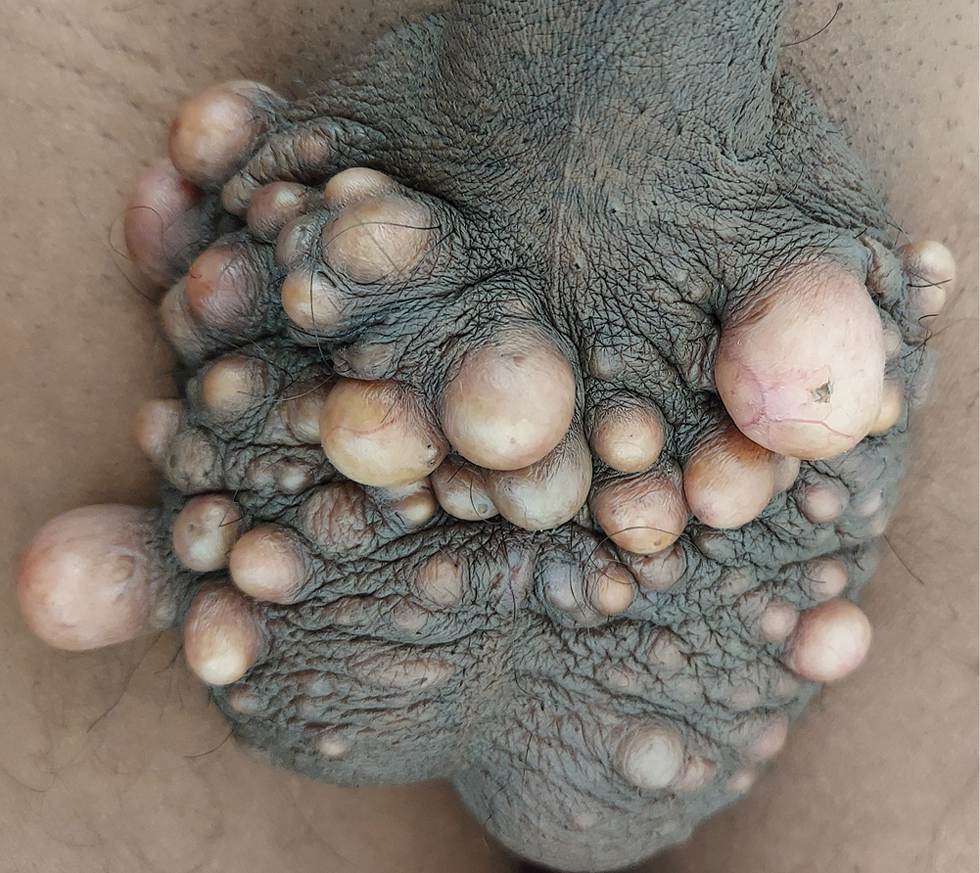
- Multiple nontender discrete and confluent, white-yellowish firm subcutaneous nodules measuring 0.2-2.5 cm in diameter attached to the scrotal skin.
Scrotal calcinosis is a rare and typically benign condition, predominantly affecting young men. The nodules are mostly asymptomatic, although some patients report itching, heaviness, and discharge of chalky material. These are not associated with any hormonal or metabolic abnormalities. The cause of the condition is debated but is likely related to the dystrophic calcification of pre-existent epidermal cysts, hair follicle cysts, or eccrine duct milia.[1] Surgical excision of the affected area and direct closure or use of flaps/grafts is generally regarded as the standard treatment.
Declaration of patient consent
Patient’s consent not required as patients identity is not disclosed or compromised.
Financial support and sponsorship
Nil.
Conflict of interest
There are no conflicts of interest.
References
- Idiopathic scrotal calcinosis: A non-elucidated pathogenesis and its surgical treatment. Rev Urol. 2011;13:95-7.
- [PubMed] [PubMed Central] [Google Scholar]


