Translate this page into:
Pigment boosting: A clue for doubtful pityriasis versicolor in the skin of color

*Corresponding author: Aravind Sivakumar, Department of Dermatology, JIPMER, Puducherry, India. aravinddermat@gmail.com
-
Received: ,
Accepted: ,
How to cite this article: Sivakumar A, Arora K. Pigment boosting: A clue for doubtful pityriasis versicolor in the skin of color. CosmoDerma 2023;3:154.
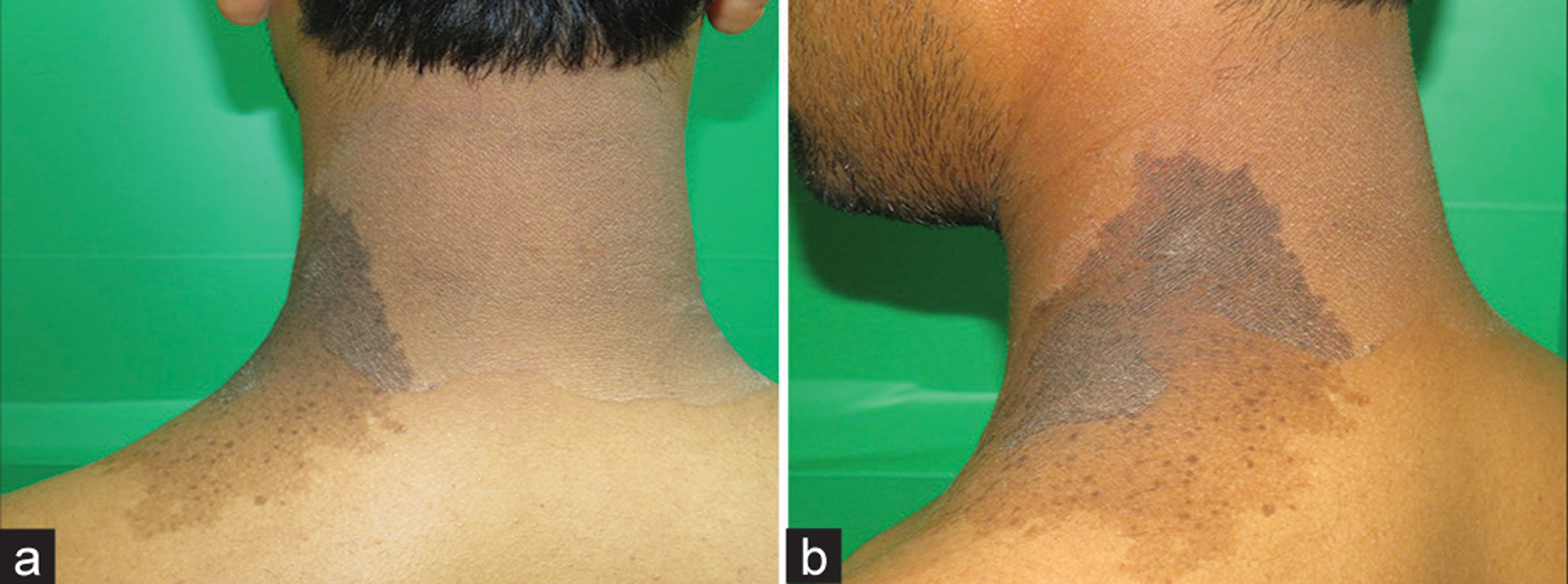
A 36-year-old male presented with asymptomatic well-defined hypopigmented and slightly erythematous macules with fine scaling present over the posterolateral aspect of the neck [Figure 1a]. He also had a nevus spilus which was adjacent to it and was overlapping with the lesion. The superimposed area consisting of both the nevi and pityriasis versicolor appeared much more pronounced and hyperpigmented compared to the lesional skin of either dermatosis [Figure 1b]. Mycological examination by potassium hydroxide mount showed spaghetti and meatballs appearance thus confirming the diagnosis of pityriasis versicolor.

- (a) Hypopigmented patch of pityriasis versicolor over the posterolateral neck along with nevus spilus. (b) The overlapping area of pityriasis versicolor and nevus spilus showing hyperpigmentation suggestive of the pigment boosting phenomenon.
Pityriasis versicolor is a superficial fungal infection caused by Malassezia species and is a common problem worldwide. It is often a diagnostic challenge, especially in skin of color due to its subtle clinical presentation and varied morphology. When present in flexural sites and confluent, it needs to be differentiated from dermatoses such as acanthosis nigricans, erythrasma, tinea corporis, and post-inflammatory hyperpigmentation. The pigment-boosting phenomenon is due to the presence of a higher density of melanocytes in the nevus and the superimposed fungal pathology further stimulates these melanocytes with the production of abnormally large melanosomes.[1,2] Hence, we describe this simple clinical vignette of how tinea versicolor can be diagnosed in the presence of such overlapping dermatosis.
Declaration of patient consent
Patient’s consent was not required as patient’s identity was not disclosed or compromised.
Conflicts of interest
There are no conflicts of interest.
Use of artificial intelligence (AI)-assisted technology for manuscript preparation
The authors confirm that there was no use of artificial intelligence (AI)-assisted technology for assisting in the writing or editing of the manuscript and no images were manipulated using AI.
Financial support and sponsorship
Nil.


