
Translate this page into:
Onychoscopy and nailfold capillaroscopy in leprosy: A case–control study

*Corresponding author: Charvi Chanana, Department of Dermatology, Venereology and Leprosy, Vardhman Mahavir Medical College and Safdarjung Hospital, New Delhi, Delhi, India. charvichanana@gmail.com
-
Received: ,
Accepted: ,
How to cite this article: Chanana C, Palanisamy M, Goyal A, Bansal S, Dash A. Onychoscopy and nail fold capillaroscopy in leprosy: A case– control study. CosmoDerma. 2024;4:28. doi: 10.25259/CSDM_260_2023
Abstract
Objectives:
Studies have reported significant number of individuals with leprosy develop nail changes of heterogeneous variety. Direct or indirect damage secondary to neural damage, repeated trauma, vascular injury, anemia or following drug therapy have been implicated. In this study, we compared nail changes in cases of leprosy with controls to determine leprosy specific nail changes through detailed onychoscopic evaluation along with nailfold capillaroscopy.
Materials and Methods:
We performed a case–control study in a tertiary care institute with 30 leprosy patients and 30 age- and sex-matched controls. The study was done over a period of three months from June 2023 to August 2023. Patients were classified as per Ridley-Jopling classification. A detailed clinical examination followed by onychoscopy and proximal nailfold (PNF) capillaroscopy using Dermlite DL4 was done in all the nails of cases and controls.
Results:
A total of 600 nails of 30 leprosy patients on treatment attending our outpatient department were studied over a span of three months. Nail changes were seen in 26 (86.6%) out of 30 patients of leprosy disease compared to 10 (33.3%) of the controls. The most common nail finding seen in leprosy patients was longitudinal melanonychia followed by leukonychia. Transverse lines, leukonychia, subungual hyperkeratosis, nail discoloration, nail pallor, onychochauxis, subungual hematoma, splinter hemorrhage, and brachyonychia were seen more in cases than in controls, and the difference was statistically significant. The PNF capillaroscopy showed microhemorrhages in eight patients of leprosy while none in controls.
Conclusion:
Nail changes seen in leprosy are multifactorial. The PNF capillaroscopy showed microhemorrhages, which could be an early feature of vascular changes in leprosy.
Keywords
Leprosy
Nail
Onychoscopy
Proximal nailfold capillaroscopy
Hansens disease

INTRODUCTION
Leprosy is a chronic, progressive infectious disease caused by Mycobacterium leprae. It primarily involves the skin and peripheral nerves. The nail changes in leprosy can be directly or indirectly related to it, neuropathy being a major cause. Other causes of nail changes include repetitive trauma, impaired blood supply, infections, vasculitic changes due to type 2 lepra reaction, and adverse effects of anti-leprosy drugs.[1]
The nail changes described are onycholysis, distorted lunula, Beau’s lines, pitting, onychorrhexis, transverse lines, leukonychia, splinter hemorrhage, dystrophy, subungual hyperkeratosis, discoloration, nail pallor, onychauxis, subungual hematoma, pterygium unguis, brachyonychia, onychogryphosis, anonychia, koilonychia, pincer nail, and melanonychia. Patients with lepromatous leprosy (LL) usually present with nail changes, which are bilaterally symmetrical and occur late. In patients with tuberculoid leprosy, these changes are unilateral, asymmetrical, and occur early.[2]
This study was conducted to compare nail changes in cases of leprosy with controls and to determine any specific nail changes in leprosy through detailed onychoscopic evaluation along with nailfold capillaroscopy. There has been a paucity of literature on proximal nailfold (PNF) capillaroscopy in leprosy patients; hence, it was done in our study.
MATERIALS AND METHODS
A case–control study was carried out in a tertiary care center in New Delhi. A total of 30 patients were studied over a period of three months. Detailed clinical examination was carried out in all the patients. Similar age- and sex-matched controls were taken. Patients were classified into Ridley–Jopling classification as tuberculoid (TT), borderline tuberculoid (BT), mid-borderline (BB), borderline lepromatous (BL), polar lepromatous (LL), and pure neuritic (PN) leprosy. Clinical photographs were taken in comparable settings of light and background. Slit skin smear and histopathological examination was done in all the cases. Demographic details were noted down. A subgroup of patients having type 1 lepra reaction and type 2 lepra reaction was made. Deformities of hands and feet were graded according to the World Health Organization guidelines. A thorough examination was carried out and nail findings were recorded
Patients having concomitant skin disease such as psoriasis and lichen planus were excluded out.
Nail changes – onycholysis, distorted lunula, Beau’s lines, pitting, onychorrhexis, transverse lines, leukonychia, splinter hemorrhage, dystrophy, subungual hyperkeratosis, discoloration, nail pallor, onychauxis, subungual hematoma, pterygium unguis, brachyonychia, onychogryphosis, anonychia, koilonychia, pincer nail, and melanonychia were looked for. A detailed onychoscopic examination was done in all the nails using DermLite DL4. Nail changes were noted down in all 20 nails. The PNF capillaroscopy changes – microhemorrhage, corkscrew capillaries, ectatic capillaries, mega capillaries, and capillary dropouts were also noted down in all the cases.
These changes were noted down in a predesigned pro forma. Clinical and onychoscopic photograph were taken. Nail changes in cases and controls were then compared and analyzed.
Scrapings for KOH examination and fungal culture were done in suspicious cases.
Ethical clearance was taken from the Ethics Committee of the hospital, and written informed consent was obtained.
RESULTS
A total of 600 nails of 30 leprosy patients on treatment attending our outpatient department were studied over a span of three months. Out of these 30 patients, 19 were male and 11 were female (Male: Female - 1.72:1). Age of the patients ranged from 20 to 65 years. Duration of the disease ranged from five months to eight years. All patients were on treatment and received multibacillary packs. Out of the 30 patients, there were 7 (23.3%) patients of BT leprosy, 1 (3.3%) of BB leprosy, 10 (33.3%) of BL leprosy, 7 (23.3%) of LL leprosy, and 5 (16.6%) of PN leprosy. Four (13%) patients presented with type 1 lepra reaction, and six (2%) patients had an episode of erythema nodosum leprosum (ENL) over the course of disease. Only two patients had active ENL as shown in Table 1.
| Nail changes | BT | BB | BL | LL | PN | ENL |
|---|---|---|---|---|---|---|
| Melanonychia | 3 | 0 | 4 | 3 | 2 | 2 |
| Discoloration | 2 | 0 | 2 | 4 | 0 | 6 |
| Nail pallor | 1 | 0 | 5 | 2 | 1 | 1 |
| Subungual hematoma | 2 | 0 | 2 | 2 | 0 | 2 |
| PNF microhemorrhage | 1 | 0 | 3 | 2 | 2 | 0 |
| Subungual hyperkeratosis | 1 | 0 | 2 | 2 | 1 | 2 |
| Splinter hemorrhage | 0 | 0 | 3 | 1 | 2 | 1 |
| Transverse lines | 0 | 0 | 1 | 2 | 1 | 2 |
| Leukonychia | 0 | 0 | 4 | 3 | 2 | 3 |
| Onychauxis | 0 | 0 | 2 | 2 | 0 | 2 |
| Brachyonychia | 0 | 0 | 1 | 1 | 2 | 1 |
| Dystrophy | 1 | 0 | 1 | 1 | 0 | 1 |
| Onychorrhexis | 1 | 0 | 1 | 0 | 0 | 0 |
| Pitting | 1 | 0 | 0 | 0 | 1 | 0 |
| Beau’s line | 1 | 1 | 0 | 0 | 0 | 0 |
| Anonychia | 0 | 0 | 0 | 1 | 0 | 1 |
| Pterygium | 0 | 0 | 1 | 0 | 0 | 0 |
| Koilonychia | 0 | 0 | 0 | 0 | 1 | 0 |
| Pincer nail | 0 | 0 | 0 | 0 | 0 | 0 |
| Ectatic capillaries | 0 | 0 | 0 | 0 | 0 | 0 |
| Mega capillaries | 0 | 0 | 0 | 0 | 0 | 0 |
| Capillary dropout | 0 | 0 | 0 | 0 | 0 | 0 |
BT: Borderline tuberculoid, BB: Mid-borderline, BL: Borderline lepromatous, LL: Polar lepromatous, PN: Pure neuritic, ENL: Erythema nodosum leprosum, PNF: Proximal nail fold
The average number of fingernails and toenails involved were 3 and 1, respectively. Fingernails were involved more than toenails.
One or more nail changes were seen in 26 (86.6%) out of 30 patients of leprosy while only 10 (33.3%) of the controls showed nail changes. The nail changes seen in cases and controls are shown in Table 2.
| Nail changes | Number of study participants showing the specific nail changes | -value | |
|---|---|---|---|
| Cases (%) | Controls (%) | ||
| Melanonychia | 14 (46.62) | 11 (36.63) | 0.432 |
| Discoloration | 14 (46.62) | 0 | 0.000 |
| Nail pallor | 10 (33.3) | 0 | 0.001 |
| Subungual hematoma | 8 (26.64) | 1 (3.33) | 0.026 |
| PNF-microhemorrhages | 8 (26.64) | 0 | 0.002 |
| Subungual hyperkeratosis | 8 (26.64) | 0 | 0.002 |
| Splinter hemorrhages | 7 (23.31) | 1 (3.33) | 0.023 |
| Transverse lines | 6 (19.98) | 0 | 0.024 |
| Leukonychia | 6 (19.98) | 0 | 0.024 |
| Onychauxis | 6 (19.98) | 0 | 0.024 |
| Brachyonychia | 5 (16.65) | 0 | 0.02 |
| Dystrophy | 4 (13.32) | 0 | 0.112 |
| Onychorrhexis | 2 (6.66) | 2 (6.66) | 1 |
| Pitting | 2 (6.66) | 1 (3.33) | 0.554 |
| Beau’s line | 2 (6.66) | 0 | 0.492 |
| Anonychia | 2 (6.66) | 0 | 0.492 |
| Pterygium | 1 (3.33) | 0 | 1 |
| Koilonychia | 1 (3.33) | 0 | 1 |
PNF: Proximal nail fold
The most common nail finding seen in leprosy patients were longitudinal melanonychia followed by leukonychia. Melanonychia was seen in 14 (46.62%) cases of leprosy and in 11 (36.63%) controls; the difference was not statistically significant. Nail changes such as transverse lines, leukonychia, subungal hyperkeratosis, nail discoloration, nail pallor, onychochauxis, subungal hematoma, splinter hemorrhage, and brachyonychia were seen more in cases than in controls with statistical significance.
Beaus’ lines, pitting, onychorrhexis, dystrophy, anonychia, and koilonychia were seen more in cases than in controls, but the difference was not statistically significant as shown in Table 2.
Nine out of ten patients with glove and stocking anesthesia showed nail changes. The most common finding seen in patients with ENL was melanonychia and leukonychia. The PNF capillaroscopy showed microhemorrhages in eight patients of leprosy while none of the controls showed any such features.
DISCUSSION
Nail changes are commonly seen in leprosy patients. Some significant nail findings were seen in cases as compared to controls. The prevalence of nail changes in leprosy patients in our study was 86.6%. Theunuo et al. reported a higher frequency (93.3%) of nail changes in their study.[3] A similar prevalence was seen by Rajput et al. (80%) while a lower prevalence in Patki and Baran (64%) and Kaur et al. (38% in paucibacillary and 68% in multibacillary cases).[2,4]
In tuberculoid pole, very few patients showed nail involvement limited to one or two nails and fingers, which had deformity either sensory or motor. Hence, nail features can be attributed to trauma, which resulted from sensory impairment. On the other hand, lepromatous pole patients showed more extensive and symmetrical nail involvement. These changes could be attributed to autonomic dysfunction, trauma, vascular changes sensory, and motor loss.
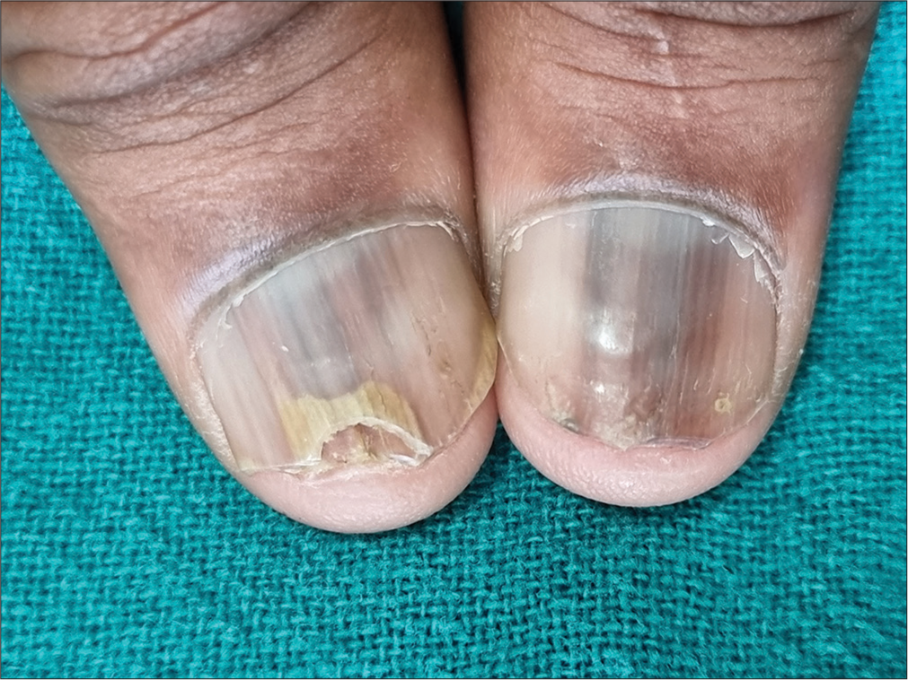
Melanonychia [Figures 1 and 2] was the most common nail finding seen in our study, which was seen more in lepromatous patients. The presence of melanonychia can be attributed to ethnicity, multidrug treatment induced or melanocyte activation due to autonomic dysfunction. Melanonychia has been the most significant finding in study by Kaur et al. and El Darouti et al.[2,5] while nail pitting was reported to be the most frequent finding by Theunuo et al. followed by longitudinal melanonychia.[3]

- Dermoscopic picture of nail showing longitudinal melanonychia (DermLite DL4, polarised mode, ×10).
- Clinical picture showing melanonychia and distal onycholysis.
In the previous studies, leukonychia has not been found to be significantly associated with leprosy since it was seen in equal number in cases and controls[2,3] while a significant association of leukonychia with leprosy was seen in our study.
Nail pallor [Figure 3], which is pale looking or light pinkish hue of the nail was seen in 10 (33.3%) cases of leprosy and none of the controls in the present study. It has been regarded as a specific feature of leprosy by some authors.[1-3] Theunuo et al. found nail pallor in 90.9% of their cases.[3] In our study, it was found more in patients with lepromatous pole, which suggests autonomic dysfunction and vascular changes to be the reason behind it.
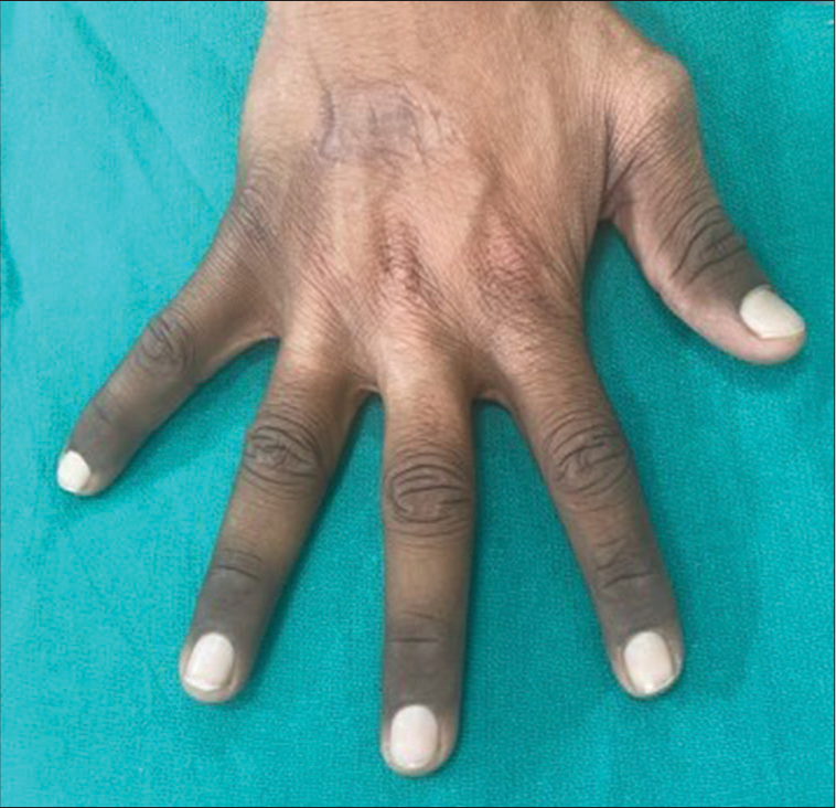
- Clinical picture showing nail pallor in all nails.
An interesting finding was seen in one of our cases. A patient of LL leprosy with nail pallor on PNF capillaroscopy showed microhemorrhages further supporting vascular changes being the causative factor behind nail pallor. Anemia due to dapsone therapy, which was regarded as a causative factor for nail pallor and was disregarded by the Theunuo et al., as they found no association with hemoglobin and nail pallor.[3]
Trophic changes such as brachyonychia (shortening of nail due to acro-osteolysis) [Figure 4], onychochauxis (thickening of the nail plate), and subungual hematoma were seen more in patients with lepromatous pole and grade 2 deformity, thereby strengthening the role of autonomic dysfunction and repeated trauma in causation of these changes. Transverse lines and Beau’s lines were also found more in cases as compared to controls and involved either one or two digits suggesting the role of repeated trauma to the germinal matrix of nail as a probable causative factor. Proximal onycholysis in a case of BT leprosy involving only index finger is shown in Figure 2. Multidrug therapy-induced changes are common. Clofazamine can cause subungual hyperkeratosis[6] while dapsone has shown Beau’s lines in some patients.[7]

- Dermoscopic picture of nail showing brachyonychia (DermLite DL4, polarized mode, ×10).
An interesting finding of half-and-half nail was found in one of our patients [Figure 5]. Other findings seen in our study were nail pitting, onychorrhexis, dystrophy, nail discoloration, anonychia, koilonychia, onychomadesis [Figure 6] altered lunula, and pterygium [Figure 7].

- (a) Clinical picture showing half-and-half nails and (b) dermoscopic picture of nail showing half-and-half nails (DermLite DL4, polarized mode, ×10).

- Clinical picture showing onychomadesis.

- Dermoscopic picture of nail showing ventral pterygium (DermLite DL4, polarized mode, ×10).
Some features such as pincer nail, ectopic nail, and paronychia were not observed in our study.
The PNF capillaroscopy has not been studied extensively in leprosy. Capillaroscopy showed us some interesting features, which were not seen by naked eyes. Eight out of 30 patients studied showed microhemorrhages. A case of nail pallor also showed microhemorrhages on capillaroscopy. None of our patients showed avascular zones, megacappilaries, corkscrew or ectatic capillaries. Lima et al. found microhemorrhages and ectatic capillaries in PNF in 40% of their patients. They did not report any avascular zones or megacapillaries.[8] Vasculitis and vasculopathy are known to occur in leprosy patients. These microhemorrhages could be an early feature of vasculopathy, which appear before nail changes.
CONCLUSION
Examination of nails is essential in leprosy patients, as they can offer important clues about the type of leprosy, potential complications of the disease, and the effect of treatment. The PNF capillaroscopy, an underutilized tool in leprosy, could detect early changes such as microhemorrhages and should be included in comprehensive evaluation of leprosy cases.
Ethical approval
Ethical clearance was taken from the Ethics Committee of the hospital. The approval date 30/3/2023.
Declaration of patient consent
The authors certify that they have obtained all appropriate patient consent.
Conflicts of interest
There are no conflicts of interest.
Use of artificial intelligence (AI)-assisted technology for manuscript preparation
The authors confirm that there was no use of artificial intelligence (AI)-assisted technology for assisting in the writing or editing of the manuscript and no images were manipulated using AI.
Financial support and sponsorship
Nil.
References
- Significance of nail changes in leprosy: A clinical review of 357 cases. Semin Dermatol. 1991;10:77-81.
- [Google Scholar]
- Nail involvement in leprosy: A study of 300 patients. Int J Lepr Other Mycobact Dis. 2003;71:320-7.
- [CrossRef] [PubMed] [Google Scholar]
- Nail changes in leprosy: Onychoscopy evaluation. Indian Dermatol Online J. 2020;11:970-4.
- [CrossRef] [PubMed] [Google Scholar]
- Nail changes in leprosy: An observational study of 125 cases. Indian Dermatol Online J. 2020;11:195-201.
- [CrossRef] [PubMed] [Google Scholar]
- Clinical study of nail changes in leprosy and comparison with nail changes in diabetic patients. J Eur Acad Dermatol Venereol. 2011;25:290-5.
- [CrossRef] [PubMed] [Google Scholar]
- Dapsone-induced erythroderma with Beau's lines. Lepr Res. 1989;60:274-7.
- [CrossRef] [Google Scholar]
- Nailfold capillaroscopy in leprosy. An Bras Dermatol. 2016;91:686-7.
- [CrossRef] [PubMed] [Google Scholar]


