
Translate this page into:
Liquid paraffin wipe: A preferred method in wet onychoscopy

*Corresponding author: Biswanath Behera, Department of Dermatology, All India Institute of Medical Sciences, Bhubaneswar, Odisha, India. biswanathbehera61@gmail.com
-
Received: ,
Accepted: ,
How to cite this article: Sangwan P, Behera B, Garg S. Liquid paraffin wipe: A preferred method in wet onychoscopy. CosmoDerma 2023;3:125.
PROBLEM
Onychoscopy, a dermoscopic examination of the nail unit, is a helpful non-invasive method to aid the diagnosis. Immersion fluid is often needed to facilitate the visualization of subsurface changes in the nail plate and nail bed.[1] Onychoscopic examination without immersion fluid hinders the visibility of dermoscopic details due to overlying scales and dirt [Figure 1a]. In contrast to dermoscopy of skin, the immersion fluid used during onychoscopy gets accumulated due to the convex surface of the nail plate. As a result, the underlying structures get blurred and hamper good dermoscopic evaluation and image acquisition. In addition, bubbles resulting from this method further impede visibility [Figure 1b]. Furthermore, the blurring effect with ultrasound gel is relatively more due to its thicker consistency, and the use of acetone for getting rid of surface dirt leaves behind white marks.
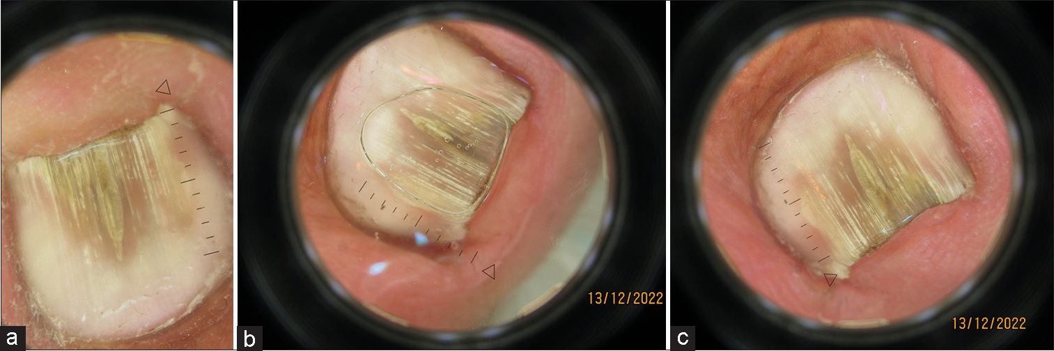
- (a) Dermoscopic image of a nail apparatus with onychomycosis showing yellow-white longitudinal streaks with the proximal reddish-brown area, blurred red streaks suggestive of splinter hemorrhage visible. Background changes and finer details are difficult to clearly delineate due to blurring and the presence of oil bubbles (after putting oil drop over the nail plate, Heine Delta 30, ×10 magnification). (b) Dermoscopic image of the same nail showing yellow-white longitudinal streaks with the proximal reddish-brown area, red streaks suggestive of splinter hemorrhage visible. Splinter hemorrhage and longitudinal streaks are clearer, with even transverse striations visible in the longitudinal streaks. Background changes and finer details have better resolution due to less blurring and the absence of any oil bubble (after putting the oil drop over the nail plate, Heine Delta 30, ×10 magnification). (c) Dermoscopic image of the same nail of onychomycosis showing yellow-white longitudinal streaks and proximal reddish-brown area which are less clear. Red streaks, which are suggestive of splinter hemorrhage and transverse striations are barely appreciable as compared to what was seen in previous images with wet onychoscopy. Background changes and finer details also have a lesser resolution. (without putting the oil drop over the nail plate, Heine Delta 30, ×10 magnification).
SOLUTION
During the liquid paraffin wipe method, we put liquid paraffin on a folded tissue paper and wipe the nail plate and folds instead of directly putting liquid paraffin drops on the nail plate surface. It facilitates the visualization of the deeper nail plate and nail bed details without interfering with the visibility [Figure 1c] due to the absence of excess oil and bubbles. In addition, it gets rid of surface dirt and scales.
Declaration of patient consent
The authors certify that they have obtained all appropriate patient consent.
Conflicts of interest
There are no conflicts of interest.
Use of artificial intelligence (AI)-assisted technology for manuscript preparation
The authors confirm that there was no use of artificial intelligence (AI)-assisted technology for assisting in the writing or editing of the manuscript and no images were manipulated using AI.
Financial support and sponsorship
Nil.
References
- Onychoscopy: A practical guide. Indian J Dermatol Venereol Leprol. 2017;83:536-49.
- [CrossRef] [PubMed] [Google Scholar]


