
Translate this page into:
Linear focal elastosis


*Corresponding author: Carol Lobo, Department of Dermatology, St. John’s Medical College, Bengaluru, Karnataka, India. carol.lobo@stjohns.in
-
Received: ,
Accepted: ,
How to cite this article: Lobo C, Kaimal S. Linear focal elastosis. CosmoDerma. 2025;5:43. doi: 10.25259/CSDM_32_2025
A 17-year-old male presented with asymptomatic streaks over the back for 6 months. There was no history of sudden weight gain or loss, use of steroids, or strenuous exercise. On examination, multiple horizontal, skin-colored, elevated, transverse bands of varying lengths were noted spanning the midline over the mid and lower back [Figure 1].
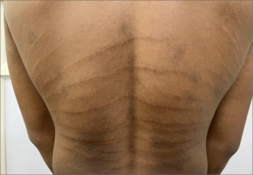
- Multiple horizontal skin-colored bands over the mid and lower back.
Based on the classical clinical findings, a diagnosis of linear focal elastosis (LFE) was made.
LFE is a rare dermal elastosis affecting males more than females and is characterized by asymptomatic yellowish palpable linear bands symmetrically distributed along the lumbosacral region.[1] The presence of atrophy and associated risk factors distinguishes striae distensae from LFE. Histopathologically, an increase in fragmented wavy elastic fibers is noted throughout the reticular dermis.[2] There is no known treatment for LFE.
Ethical approval
Institutional Review Board approval is not required.
Declaration of patient consent
The authors certify that they have obtained all appropriate patient consent.
Conflicts of interest
There are no conflicts of interest.
Use of artificial intelligence (AI)-assisted technology for manuscript preparation
The authors confirm that there was no use of artificial intelligence (AI)-assisted technology for assisting in the writing or editing of the manuscript and no images were manipulated using AI.
Financial support and sponsorship: Nil.
References
- Acquired disorders of elastic tissue: Part I. increased elastic tissue and solar elastotic syndromes. J Am Acad Dermatol. 2004;51:1-21.
- [CrossRef] [PubMed] [Google Scholar]
- Disorders of elastic tissue In: Skin pathology (2nd ed). London, England: Churchill Livingstone; 2002. p. :381-404.
- [Google Scholar]


