Translate this page into:
Florid cutaneous neurofibromas in neurofibromatosis type 1: A classic presentation

*Corresponding author: Manju Daroach, Department of Dermatology, AIIMS, Bilaspur, Himachal Pradesh, India. daroachmanju@gmail.com
-
Received: ,
Accepted: ,
How to cite this article: Chauhan P, Bansal V, Daroach M. Florid cutaneous neurofibromas in neurofibromatosis type 1: A classic presentation. CosmoDerma. 2024;4:31. doi: 10.25259/CSDM_16_2024
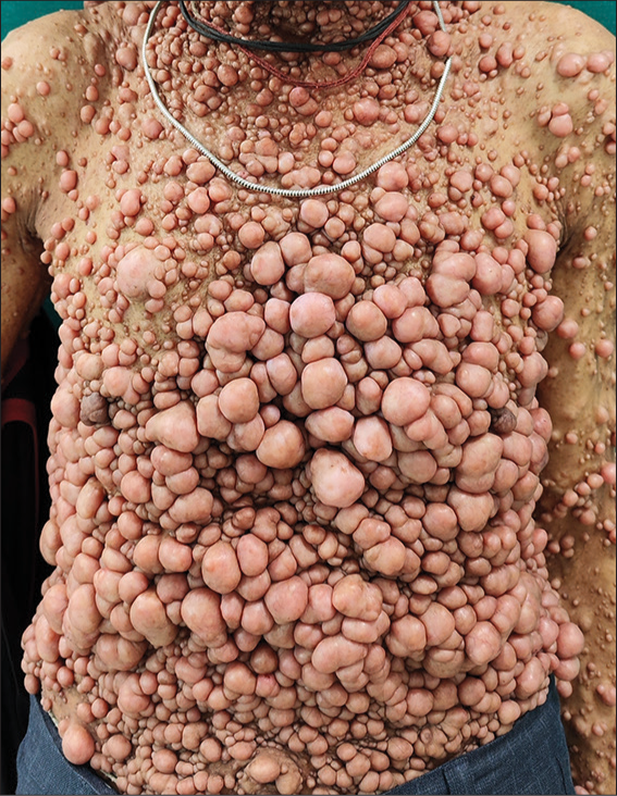
A 40-year-old man presented to the dermatology outpatient department with multiple nodular swellings over his body since childhood, which were progressively increasing in size and number. His mother also had similar lesions. On examination, uncountable soft skin-colored nodular swellings were present all over the body [Figure 1]. There was freckle-like pigmentation evident all over his body including axillae and palms. Furthermore, multiple café au lait macules were present over the trunk and limbs. On ophthalmologic examination, Lisch nodules were seen on the iris. No skeletal abnormalities were found on orthopedic examination. A final diagnosis of neurofibromatosis type 1 (NF-1) was made according to the revised diagnostic criteria for NF-1.[1] The patient was counseled regarding the nature of the disease and management options for a neurofibroma.

- Patient of Von Recklinghausen’s disease with florid cutaneous neurofibromas and freckle-like pigmentation.
Von Recklinghausen’s NF-1 is one of the most common autosomal-dominant neurocutaneous conditions in humans characterized by multiple soft-tissue tumors arising from Schwann cells and fibroblasts caused by a mutation in the neurofibromin one gene located on chromosome 17q11.2.1.[2] The hallmark lesion is the neurofibroma, a benign peripheral nerve sheath tumor, with subtypes: Cutaneous, subcutaneous, nodular or diffuse plexiform, and spinal. The earliest clinical manifestation of NF-1 is café-au-lait macules, axillary and inguinal freckling is another common clinical feature of NF-1 and is usually detected in affected individuals by age 5–8 years, Lisch nodules are benign melanocytic hamartomas of the iris, typically first noticed in children aged 5–10 years.[3] Management of neurofibromas is challenging. There is no effective medical treatment available. Physical removal has been the mainstay of therapy and gives excellent cosmetic results.
Our patient had a very typical case of NF-1, which presents considerable interest because of the florid neurofibromas over the skin. Such cases require a detailed history and examination to rule out internal organ involvement. These patients may be treated by surgery for cosmetic improvement and also genetic counseling is necessary.
Ethical approval
Institutional Review Board approval is not required.
Declaration of patient consent
The authors certify that they have obtained all appropriate patient consent.
Conflicts of interest
There are no conflicts of interest.
Use of artificial intelligence (AI)-assisted technology for manuscript preparation
The authors confirm that there was no use of artificial intelligence (AI)-assisted technology for assisting in the writing or editing of the manuscript and no images were manipulated using AI.
Financial support and sponsorship
Nil.
References
- Revised diagnostic criteria for neurofibromatosis type 1 and Legius syndrome: An international consensus recommendation. Genet Med. 2021;23:1506-13.
- [CrossRef] [PubMed] [Google Scholar]
- Coexistence of neurofibromatosis 1 and vitiligo. Cosmoderma. 2023;3:126.
- [CrossRef] [Google Scholar]
- Neurofibromatosis type 1: A multidisciplinary approach to care. Lancet Neurol. 2014;13:834-43.
- [CrossRef] [PubMed] [Google Scholar]


