Translate this page into:
A man with scars, ulcers, and pigmentation

*Corresponding author: Anupam Das, Department of Dermatology, KPC Medical College and Hospital, Kolkata, West Bengal, India. anupamdasdr@gmail.com
-
Received: ,
Accepted: ,
How to cite this article: Kumar P, Das A. A man with scars, ulcers, and pigmentation. CosmoDerma 2022;2:32.
CASE HISTORY, EXAMINATION, AND INVESTIGATION FINDINGS
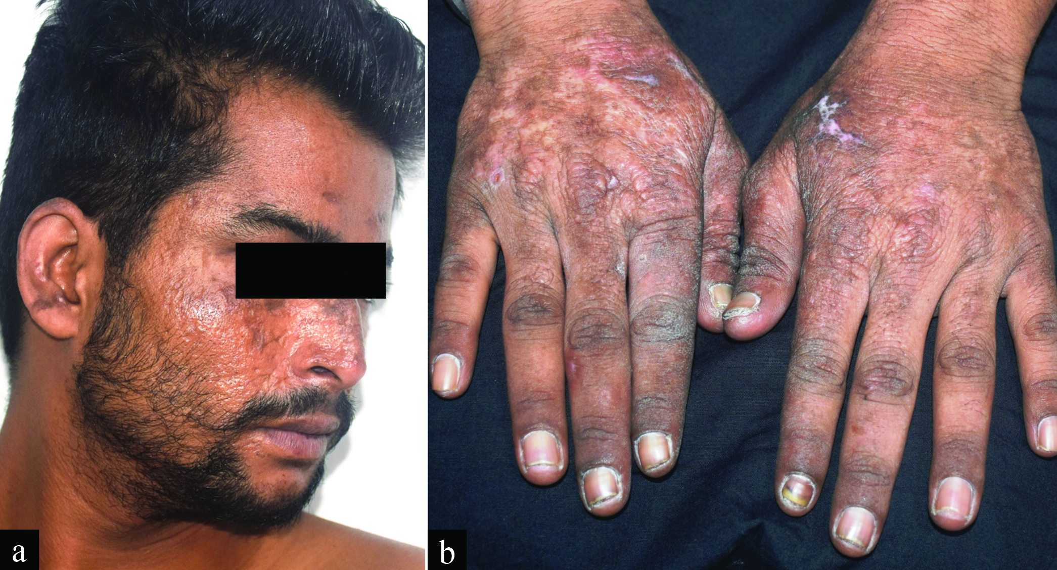
A 21-year-old man presented with recurrent sunburns, and ulcers over the face and hands, present for 13 years. Four out of six siblings had similar complaints. There was a history of erythema, burning, and ulceration on sun exposure; healing with atrophic scarring and reticulate pigmentation. Examination revealed atrophic scarring and dyspigmentation, over the face, ears, and dorsum of hands. Crusted ulcers were noted on the ear and dorsum of hands [Figure 1]. Laboratory investigations revealed hemoglobin 10.7 gm/dl, free protoporphyrin 24110.9 nMol/L (relative amount 96.72%), total protoporphyrin 24928.07 nMol/L, and zinc protoporphyrin 123.07 μMol/L. The liver profile revealed elevated liver enzymes (SGPT 108 U/L, SGOT 96 U/L). Genetic study could not be done due to logistic reasons.

- Atrophic scarring and dyspigmentation over the face and dorsum of hands. Note the presence of crusted ulcers over the ear and dorsum of left hand.
What is the diagnosis?
Answer:
Erythropoietic protoporphyria
DISCUSSION
Elevated free protoporphyrin with mildly increased zinc protoporphyrin, suggested a diagnosis of erythropoietic protoporphyria (EPP). Our patient has been counseled to avoid sunlight and apply sunscreens. He has been asked to attend our clinic for periodic follow-up to rule out systemic iron overload and liver disease.
Erythropoietic protoporphyria (EPP) is a photodermatosis with autosomal recessive inheritance, characterized by recurrent painful sunburns with rare vesiculation or blistering. Most commonly affected sites are face ear, and dorsum of hands. The severity of the condition may vary—some patients may develop painful sunburn with seconds of sun exposure while some others may require hours of sun exposure to develop sunburn. Sunburns may persist for hours to days depending on the intensity and duration of sun exposure. Associated photo-onycholysis may develop. Chronic cases go on to develop scarring, thickened leathery skin, and focal hyperkeratosis. Milia or hypertrichosis, seen in other cutaneous porphyrias, are absent. Rarely, EPP may be seen in patients with myelodysplasia or myeloid leukemia where chromosomal instability may lead to loss of part of chromosome 18. Deficient activity of the enzyme ferrochelatase leads to the accumulation of free protoporphyrins in the hematopoietic and hepatobiliary systems. Recurrent photo-exposures lead to the development of lichenification, leathery pseudovesicles, perioral grooving, and loss of lunulae of the nails.[1] About 20-30% of individuals present with mild elevations of liver enzymes, and 5% of the patients may land up into severe hepatic dysfunction.[2] Increasing hepatic dysfunction is associated with aggravation of photosensitivity. Diagnosis of EPP is done by detection of markedly elevated levels of free erythrocyte protoporphyrin and/or by identifying mutations in gene encoding ferrochelatase.[3]
Management of the disease depends on the symptoms, chronicity, and systemic involvement. Afamelanotide has been approved for acute photosensitivity.[4] Photoprotection is the cornerstone of therapy. Hepatic manifestations are managed with cholestyramine, plasmapheresis, intravenous haemin, and liver transplantation. Iron replacement therapy is found to provide good results. Patients should be routinely assessed for erythrocyte protoporphyrin levels, blood parameters, hepatobiliary profile, and vitamin D levels.
Declaration of patient consent
The authors certify that they have obtained all appropriate patient consent.
Financial support and sponsorship
Nil.
Conflicts of interest
There are no conflicts of interest.
References
- Clinical, biochemical, and genetic characterization of North American patients with erythropoietin protoporphyria and X-linked protoporphyria. JAMA Dermatol. 2017;153:789-96.
- [CrossRef] [PubMed] [Google Scholar]
- Gene dosage analysis identifies large deletions of the FECH gene in 10% of families with erythropoietin protoporphyria. J Invest Dermatol. 2007;127:2790-4.
- [CrossRef] [PubMed] [Google Scholar]
- Long-term observational study of afamelanotide in 115 patients with erythropoietic protoporphyria. Br J Dermatol. 2015;172:1601-12.
- [CrossRef] [PubMed] [Google Scholar]


