
Translate this page into:
Systematized nevus lipomatosus

*Corresponding author: Neetu Bhari, Department of Dermatology and Venereology, All India Institute of Medical Sciences, New Delhi, India. drntbhari@gmail.com
-
Received: ,
Accepted: ,
How to cite this article: Yadav P, Sharma A, Bhari N. Systematized nevus lipomatosus. CosmoDerma. 2025;5:60. doi: 10.25259/CSDM_53_2025
A 13-year-old female presented with gradually progressive asymptomatic skin-colored to hyperpigmented soft plaques with lobulated surface on the back, neck, and face in Blaschko-linear pattern for the past 5 years [Figure 1a and b]. For the lesions on the back, the clinical differential diagnoses included connective tissue nevus, nevus lipomatosus, and epidermal nevus. For the facial lesions, the differentials considered were connective tissue nevus, nevus lipomatosus, and hemihypertrophy. Histopathology from the lower back lesion revealed mild hyperkeratosis, acanthosis, and aggregates of mature adipocytes interspersed between collagen bundles in the reticular dermis, not connected to the subcutaneous fat [Figure 2], suggestive of nevus lipomatosus cutaneous superficialis. Nevus lipomatosus is a hamartoma of adipocytes that present as painless pedunculated skin-colored to yellowish nodules in a zosteriform distribution with a predilection for the pelvic girdle.[1] Our case had atypical involvement of the face along with extensive Blaschko-linear involvement akin to the systematized distribution described for epidermal nevi.[2]
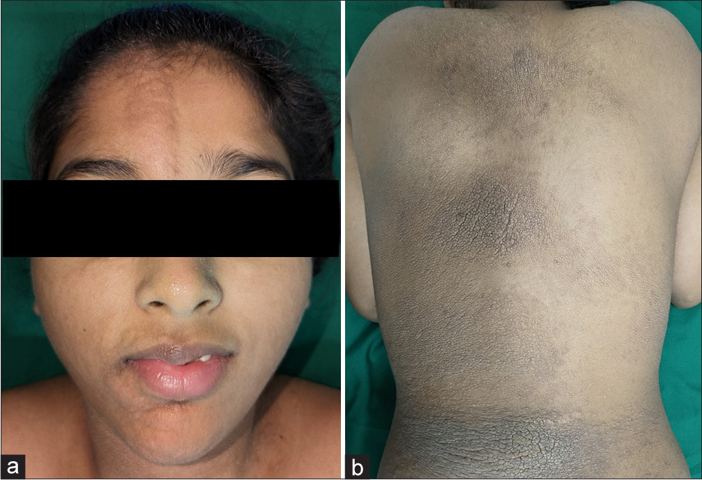
- (a) Nevus lipomatosus involving the right side of the face as soft, barely elevated skin-colored, ill-defined plaques with sharp midline demarcation, causing facial asymmetry, (b) Nevus lipomatosus involving the right side of the back as hyperpigmented cobblestone plaques, extending to the contralateral side as multiple linear lesions.
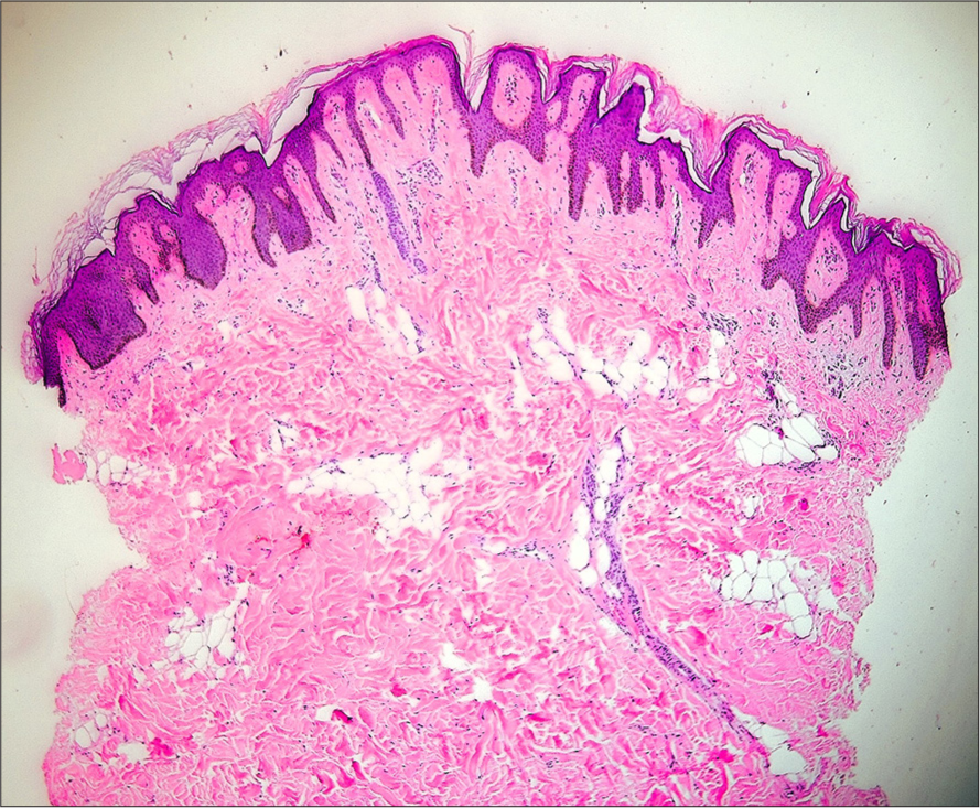
- Histopathology of nevus lipomatosus showing mild hyperkeratosis, papillomatosis, epidermal acanthosis, and the presence of multiple mature adipocytes in reticular dermis with no connection to subcutaneous fat (hematoxylin and eosin, ×40).
Ethical approval:
Institutional Review Board approval is not required.
Declaration of patient consent:
The authors certify that they have obtained all appropriate patient consent.
Conflicts of interest:
There are no conflicts of interest.
Use of artificial intelligence (AI)-assisted technology for manuscript preparation:
The authors confirm that there was no use of artificial intelligence (AI)-assisted technology for assisting in the writing or editing of the manuscript and no images were manipulated using AI.
Financial support and sponsorship: Nil.
References
- Nevus lipomatosus superficialis: A rare cutaneous hamartoma. Indian Dermatol Online J. 2014;5:109-10.
- [CrossRef] [PubMed] [Google Scholar]
- Tumours and cysts of the epidermis In: Elder DE, ed. Lever’s histopathology of the skin (10th ed). Philadelphia, PA: Wolters Kluwer/Lippincott Williams and Williams; 2008. p. :791.
- [Google Scholar]


