
Translate this page into:
Cut kiwi fruit sign in acne excoriee


*Corresponding author: Sohel Shabbir Inamdar, Department of Dermatology, Pimpri Chinchwad Municipal Coroporation’s Post Graduate Institute, Yashwantrao Chavan Memorial Hospital, Pune, Maharashtra, India. sohelinamdar1998@gmail.com
-
Received: ,
Accepted: ,
How to cite this article: Inamdar SS, Palaskar NM. Cut kiwi fruit sign in acne excoriee. CosmoDerma. 2025;5:55. doi: 10.25259/CSDM_51_2025
A 15-year-old male came to the Outpatient Department with complaints of multiple papules over the face with itching for one year and aggravated since the past month with the onset of the board examination [Figure 1]. On examination, there were multiple skin colored papules of size 3–5 mm, discretely present all over the face, predominantly over the forehead, and a few excoriation marks with hyperpigmentation, along with multiple plugs of keratin and sebum in dilated pilosebaceous orifices were seen. The diagnosis of acne excoriee was made on the basis of history and clinical examination [Figure 2]. On dermoscopic examination of papular lesions using a DermLite DL3N dermoscope with ×10 magnification in polarized mode, one of the papules showed three distinct zones with an innermost zone of hypopigmentation, a middle zone of light brown hyperpigmentation with radial white colored streaks, and an outer zone of rim of hypopigmentation. The overall appearance of the lesion resembles the “cut section of a kiwi fruit.” Cut kiwi fruit sign is earlier reported in condition like Mudi-chood Dermatosis. It occurs due central hypopigmentation and excoriation from center of the lesion to the periphery leading to serous discharge accumulation in it and drying of that discharge gives radial streak pattern with surrounding halo rim of hypopigmentation. This is 1st time we are reporting cut kiwi fruit sign in acne excoriee on dermoscopy.
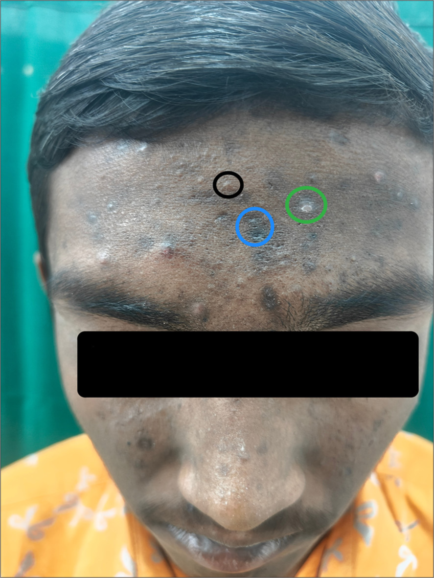
- On clinical examination, multiple skin-colored papules (black circle) with few excoriation marks (green circle) and hyperpigmentation (blue circle) are predominantly seen over the forehead.
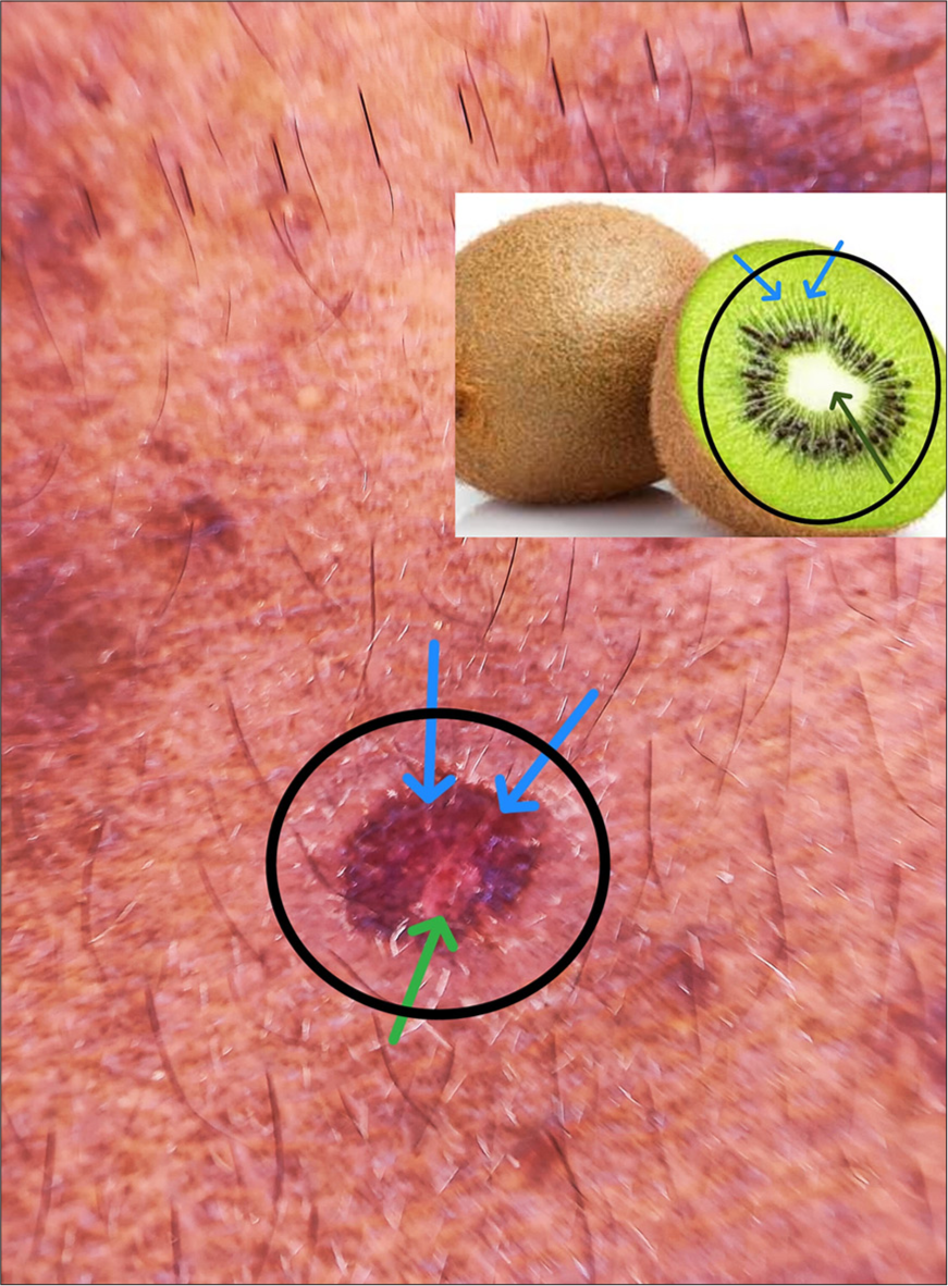
- Dermoscopic examination showing, three distinct zones with an innermost zone of hypopigmentation (green arrow), middle zone of light brown hyperpigmentation with radial white colored streaks (blue arrow), and an outer zone of rim of hypopigmentation (black circle) giving rise to an appearance resembling a cut section of kiwi fruit. Inset image showing cut section of kiwi fruit with central white area (black arrow) and radiating white streaks (blue arrow).
Ethical approval:
Institutional Review Board approval is not required.
Declaration of patient consent:
The authors certify that they have obtained all appropriate patient consent.
Conflicts of interest:
There are no conflicts of interest.
Use of artificial intelligence (AI)-assisted technology for manuscript preparation:
The authors confirm that there was no use of artificial intelligence (AI)-assisted technology for assisting in the writing or editing of the manuscript and no images were manipulated using AI.
Financial support and sponsorship: Nil.


