
Translate this page into:
Fibrous cephalic plaque of tuberous sclerosis complex

*Corresponding author: Suvesh Singh, Department of Dermatology and STD, All India Institute of Medical Sciences, Patna, Bihar, India. suveshsingh2658@gmail.com
-
Received: ,
Accepted: ,
How to cite this article: Paul D, Snehal A, Singh S. Fibrous cephalic plaque of tuberous sclerosis complex. CosmoDerma. 2024;4:47. doi: 10.25259/CSDM_33_2024
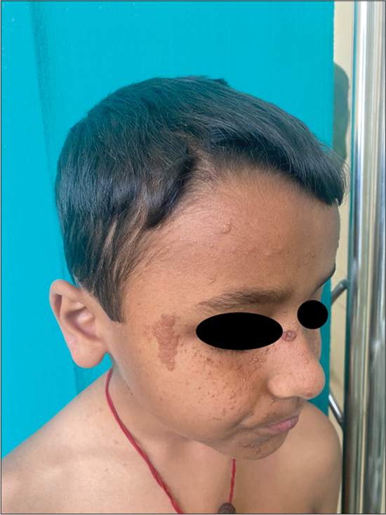
A 9-year-old boy presented with multiple reddish to skin-colored discrete papules over his face for four years. Six months later, the patient developed two skin-colored elevated lesions over the left forehead and right temple. His mother also gave a history of multiple episodes of generalized tonic-clonic seizures for two years and is currently on anti-epileptic medications. On examination, multiple angiofibromas were noted over the face. Two well-defined skin-colored plaques of size 5 × 4 cm and 4 × 2 cm were present over the left forehead and the right temple suggestive of fibrous cephalic plaque (FCP) [Figures 1 and 2]. On examination of the trunk, an irregularly shaped, thickened, and slightly elevated skin-colored patch of size 5 × 3 cm was noted as suggestive of a shagreen patch [Figure 3]. A diagnosis of tuberous sclerosis complex (TSC) was made. An ultrasound whole abdomen was performed, which revealed normal findings, and a magnetic resonance imaging brain was performed, but reports are awaited. The patient was referred to the pediatric department for further evaluation. The TSC is an autosomal dominant neurocutaneous disorder.[1] It is characterized by angiofibromas, FCP, hypopigmented macules, shagreen patches, and Koenen tumors.[2] Loss of function mutation in the tumor suppressor gene TSC1 or TSC2 results in increased activity of the mammalian target of rapamycin that causes aberrant cell proliferation and differentiation, leading to the formation of hamartomas.[2] The FCP clinically presents as skin-colored to brownish plaque with a rubbery to firm consistency.[3] They are commonly located over the forehead, face, neck, and scalp with sizes ranging from 1 cm to 5 cm.[2] The FCP may be present at birth or appear in early childhood.[2] The histopathological study from the FCP showed thickened, disorganized bundles of reticular collagen in the dermis with decreased elastic fibers compared to normal skin.[3] In our patient, the presence of two major features, that is, facial angiofibroma/FCP and shagreen patch clinched the diagnosis as per the updated diagnostic criteria for TSC 2012.[4] Biopsy findings were consistent with FCP from the forehead plaque [Figure 4]. The patient was started on sirolimus 0.1% ointment for angiofibroma but has yet to follow-up after the initial visit.

- Fibrous cephalic plaque over the forehead with multiple angiofibromas over the left side of the face.
- Fibrous cephalic plaque over the right temple region with multiple angiofibromas over the right side of the face.
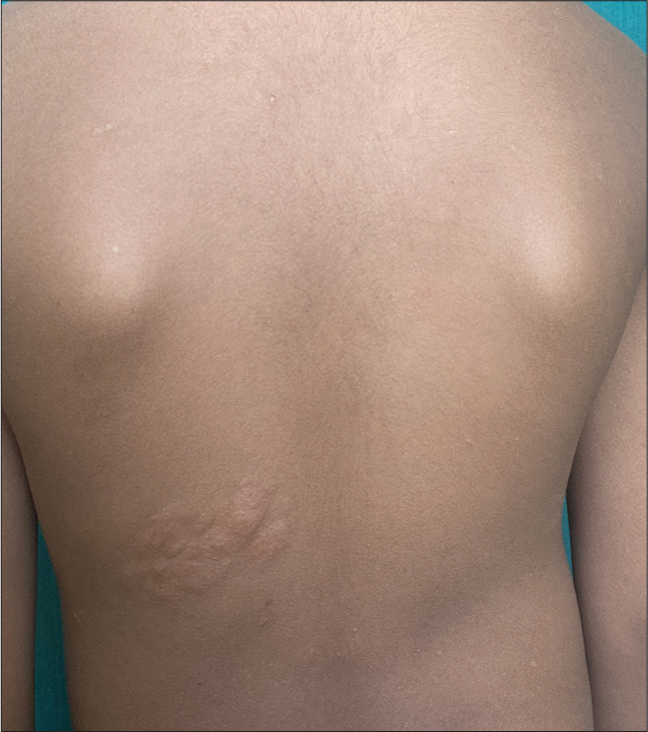
- Shagreen patch over left mid-lower back.
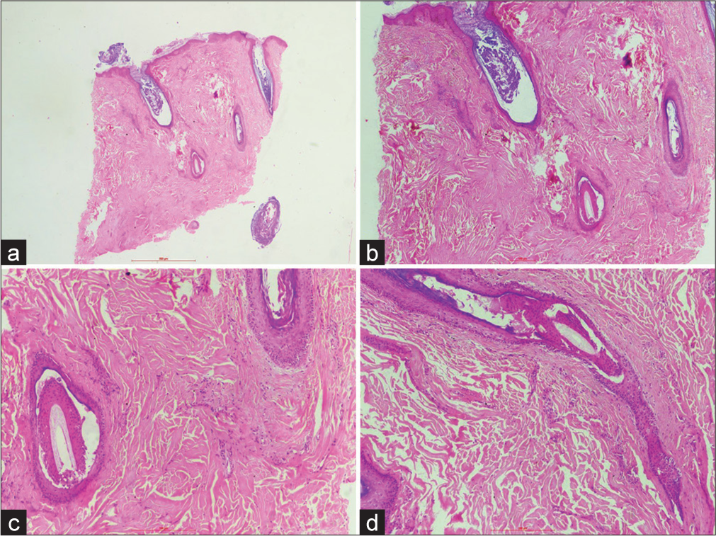
- (a) Skin biopsy shows basket-weave orthokeratosis with the presence of keratotic plugs. (H&E; ×20) (b) The dermis shows irregularly arranged thick bundles of reticular collagen bundles (H&E; ×40), (c) The involvement of hair follicles leads to perifollicular fibrosis (H&E; ×100), (d) The hair follicles are distorted due to irregularly thickened collagen (H&E; ×100).
Ethical approval
The Institutional Review Board approval is not required.
Declaration of patient consent
The authors certify that they have obtained all appropriate patient consent.
Conflicts of interest
There are no conflicts of interest.
Use of artificial intelligence (AI)-assisted technology for manuscript preparation
The authors confirm that there was no use of artificial intelligence (AI)-assisted technology for assisting in the writing or editing of the manuscript and no images were manipulated using AI.
Financial support and sponsorship
Nil.
References
- Cephalic fibrous plaque in tuberous sclerosis. Indian Pediatr. 2023;60:327.
- [CrossRef] [PubMed] [Google Scholar]
- Histopathological features of fibrous cephalic plaques in tuberous sclerosis complex. Histopathology. 2021;79:619-28.
- [CrossRef] [PubMed] [Google Scholar]
- Fibrous cephalic plaques in tuberous sclerosis complex. J Am Acad Dermatol. 2018;78:717-24.
- [CrossRef] [PubMed] [Google Scholar]
- Tuberous sclerosis complex diagnostic criteria update: Recommendations of the 2012 International Tuberous Sclerosis Complex Consensus Conference. Pediatr Neurol. 2013;49:243-54.
- [CrossRef] [PubMed] [Google Scholar]


