
Translate this page into:
Transillumination: A simple cost-effective diagnostic tool for subungual glomus tumor


*Corresponding author: Vishal Gaurav, Department of Dermatology and Venereology, Maulana Azad Medical College, New Delhi, India. mevishalgaurav@gmail.com
-
Received: ,
Accepted: ,
How to cite this article: Patel V, Gaurav V. Transillumination: A simple cost-effective diagnostic tool for subungual glomus tumor. CosmoDerma. 2024;4:46. doi: 10.25259/CSDM_41_2024
PROBLEM
A 49-year-old man presented to the dermatology outpatient department with pain in his right ring finger for three years. The pain was intense, particularly during the winter months and was also provoked by the slightest trauma, often radiating to the right shoulder. On examination, there was a small erythematous to bluish mass visible through the distal nail plate [Figure 1]. There was sharp pain upon pressure without any nail deformity. Transillumination clearly demarcated the tumor from the surrounding nail bed [Figure 2]. Dermoscopy showed a structureless purplish to reddish subungual spot and minimal onycholysis [Figure 3]. Magnetic resonance imaging showed subtle short tau inversion recovery hyperintensity in the nail bed region of the 4th digit measuring 5 × 6 mm [Figure 4]. The patient was diagnosed with ungual glomus tumor involving the right ring finger and underwent partial nail plate avulsion followed by surgical removal of the tumor.

- Small erythematous to bluish mass visible through the distal nail plate.
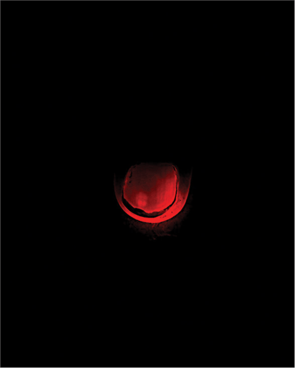
- Transillumination clearly demarcated the tumor.

- Structureless purplish to reddish subungual spot on dermoscopy (Illuco IDS 1100, Polarized, ×10).
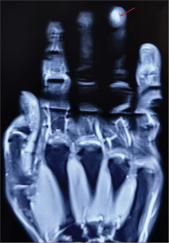
- Short tau inversion recovery hyperintensity (red arrow) on T2-weighted magnetic resonance imaging with gadolinium contrast in the nail bed region of the right ring finger.
SOLUTION
Given the challenges faced in diagnosing subungual glomus tumors, especially with limitations in imaging modalities such as magnetic resonance imaging due to cost and accessibility, transillumination emerges as a promising solution. Fingertip transillumination, a non-invasive and cost-effective technique, illuminates the subungual area with a single, narrow beam of light in a dark room. This method aids in visualizing subungual glomus tumors by delineating them from the surrounding normal tissue. Incorporating transillumination as a diagnostic tool facilitates accurate diagnosis and pre-operative localization. This approach not only enhances clinical assessment but also minimizes the need for costly or inaccessible imaging modalities, ensuring efficient management of subungual glomus tumors.[1]
Ethical approval
Institutional Review Board approval is not required.
Declaration of patient consent
The authors certify that they have obtained all appropriate patient consent.
Conflicts of interest
There are no conflicts of interest.
Use of artificial intelligence (AI)-assisted technology for manuscript preparation
The authors confirm that there was no use of artificial intelligence (AI)-assisted technology for assisting in the writing or editing of the manuscript and no images were manipulated using AI.
Financial support and sponsorship
Nil.
References
- Transillumination: A diagnostic tool to assess subungual glomus tumors. Skin Appendage Disord. 2021;7:231-3.
- [CrossRef] [PubMed] [Google Scholar]


