Translate this page into:
Halo phenomenon around hemangioma in a neonate

*Corresponding author: Arun Somasundaram, Department of Dermatology, Jawaharlal Institute of Postgraduate Medical Education and Research, Puducherry, India. arunsomasundaram25@gmail.com
-
Received: ,
Accepted: ,
How to cite this article: Somasundaram A. Halo phenomenon around hemangioma in a neonate. CosmoDerma 2023;3:127.
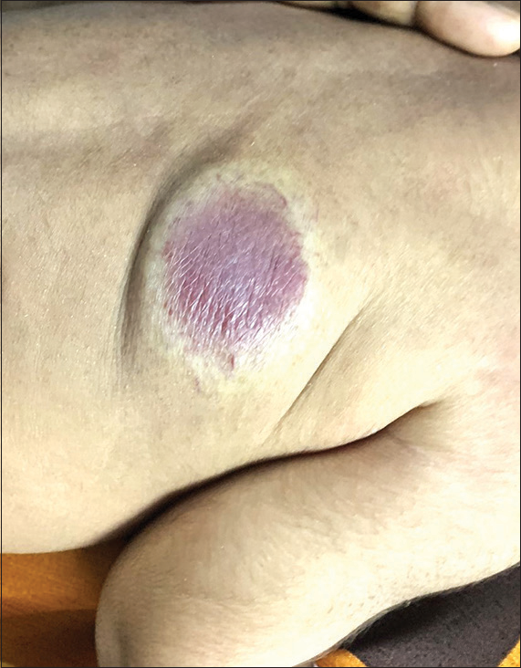
One-day-old neonate was referred from the neonatal intensive care unit for skin lesion over the back present since birth. The neonate was born of a non-consanguineous parent, a full-term baby delivered by cesarean section, and the indication was prolonged labor in the mother. On examination, there was a dull red plaque with a minimal bluish hue of size 3×2.5 cm with a surrounding halo-like rim of pallor and surface telangiectasias [Figure 1]. The rest of the cutaneous examination was noncontributory. Based on the history and clinical examination, a diagnosis of congenital hemangioma was made and the mother was asked to follow-up for the progression of the hemangioma. Infantile hemangioma was ruled out as they usually present at birth as flat telangiectatic macule rather than a fully evolved vascular tumor.

- A 1-day-old neonate with a dull red plaque of size 3×2.5 cm with a peripheral halo-like rim of pallor and surface telangiectasias suggestive of congenital hemangioma.
The halo phenomenon is observed in conditions such as halo nevus, blue nevus, Spitz nevus, psoriasis, warts, molluscum contagiosum, and lichen planus. Congenital hemangiomas can have a halo-like rim of pallor at birth giving a clue to the diagnosis. They are uncommon benign vascular tumors fully formed at birth and do not exhibit postnatal proliferation. Three main types include rapidly involuting congenital hemangioma, non-involuting congenital hemangioma, and partially involuting congenital hemangioma. Activating mutations in GNAQ and GNA11 cause upregulation of the MAPK pathway resulting in proliferation.[1] Morphology includes plaques covered with coarse telangiectasias and violaceous nodules with a halo-like rim of pallor on the adjacent skin.[1] Transient coagulopathy and thrombocytopenia are reported in the literature. The involution starts days to weeks after birth and is complete in 6–14 months leaving behind redundant atrophic hypopigmented skin in the case of rapidly involuting type while non-involuting persists with age. Diagnosis is purely clinical and imaging may be needed to delineate extent if necessary.
Declaration of patient consent
Patient’s consent not required as patients identity is not disclosed or compromised.
Conflicts of interest
There are no conflicts of interest.
Use of artificial intelligence (AI)-assisted technology for manuscript preparation
The authors confirm that there was no use of artificial intelligence (AI)-assisted technology for assisting in the writing or editing of the manuscript and no images were manipulated using AI.
Financial support and sponsorship
Nil.
References
- Congenital hemangioma: A Case report of a finding every physician should know. Cureus. 2018;10:e2485.
- [CrossRef] [Google Scholar]


