
Translate this page into:
Telangiectatic mats in systemic sclerosis

*Corresponding author: Manju Daroach, Department of Dermatology, All India Institute of Medical Sciences, Bilaspur, Chhattisgarh, India. daroachmanju@gmail.comm
-
Received: ,
Accepted: ,
How to cite this article: Dhiman A, Daroach M. Telangiectatic mats in systemic sclerosis. CosmoDerma. 2025;5:26. doi: 10.25259/CSDM_3_2025
A 54-year-old female known case of proximal systemic sclerosis (having skin thickening proximal to elbows, knees, and trunk, digital pitted scars, Raynaud’s history with positive anti-topoisomerase antibodies) for five years referred from the rheumatology outpatient department with multiple reddish lesions over the face and palms for the past five months. On examination, there were multiple mat-like telangiectasias over the face [Figure 1a and b] and bilateral palms [Figure 1c]. Telangiectatic mats or mat-like telangiectasia, characterized by flat, macular clusters of uniform small vessels around 0.5 cm in size, usually appear in rectangular formations. These are most often observed on the face, lips, palms, and the backs of the hands and can also be found around the lips, tongue, and mucous membranes. The presence of cutaneous telangiectasias indicates ongoing vascular damage and may act as a useful clinical biomarker for systemic vascular abnormalities, including pulmonary vascular disease, which has the potential to progress to pulmonary arterial hypertension (PAH). A higher number of telangiectasia is associated with an increased risk of PAH in patients with scleroderma.[1]
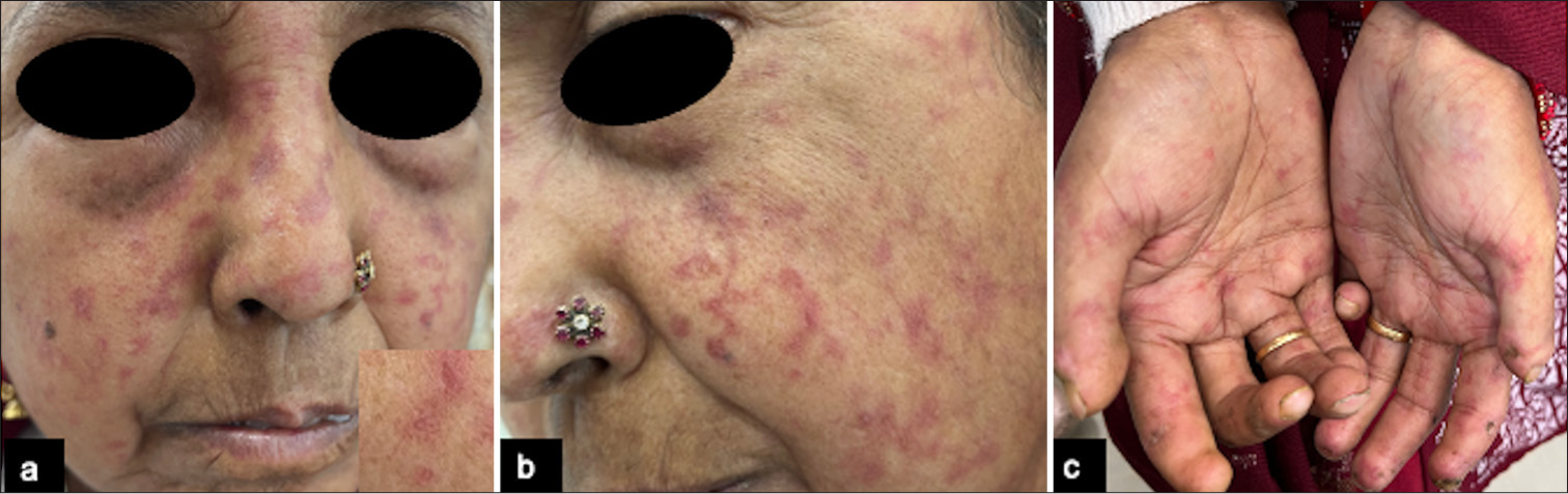
- (a) Mat-like telangiectasia over the face, inset showing dilated capillary. (b) Mat-like telangiectasia over the left side of the face. (c) Mat-like telangiectasia over palms bilaterally.
Ethical approval
Institutional Review Board approval is not required.
Declaration of patient consent
The authors certify that they have obtained all appropriate patient consent.
Conflicts of interest
There are no conflicts of interest.
Use of artificial intelligence (AI)-assisted technology for manuscript preparation
The authors confirm that there was no use of artificial intelligence (AI)-assisted technology for assisting in the writing or editing of the manuscript and no images were manipulated using AI.
Financial support and sponsorship: Nil.
References
- Telangiectases in scleroderma: A potential clinical marker of pulmonary arterial hypertension. J Rheumatol. 2010;37:98-104.
- [CrossRef] [PubMed] [Google Scholar]


