
Translate this page into:
Skin-colored nodule on the forehead with telangiectasia

*Corresponding author: Pradeep S. Nair, Department of Dermatology and Venereology, Government Medical College, Thiruvananthapuram, Kerala, India. dvmchtvm@yahoo.co.in
-
Received: ,
Accepted: ,
How to cite this article: Nair PS. Skin-colored nodule on the forehead with telangiectasia. CosmoDerma. 2024;4:61. doi: 10.25259/CSDM_29_2024
A 68-year-old male presented with a nodule on the forehead of seven months’ duration. The lesion first started as a skin-colored papule, was tender, and attained the present size within a span of seven months. There was no history of ulceration. On examination, there was a well-defined skin-colored nodule of 2 × 2.5 cm on the forehead above the right eyebrow [Figure 1]. The surface showed telangiectasia, and the nodule was tender on palpation. There was no regional lymphadenopathy. Dermoscopy showed red areas with dilated blood vessels. An excision biopsy was done. Biopsy showed the dermis to be packed with tumor cells arranged in sheets and nests and tubular lumina filled with colloid material. High power showed two types of cells: Small cells with clear cytoplasm and tiny basophilic nuclei [Figure 2] and fusiform cells with basophilic cytoplasm [Figure 3], diagnostic of nodular hidradenoma (NH). The NA also known as clear cell hidradenoma is a benign tumor of eccrine differentiation. The tumor most commonly affects the head and neck region and is sometimes a painful tumor. The tumor cells express epithelial membrane antigen and carcinoembryonic antigen. Malignant transformation is rare but can be aggressive. The NH is known for frequent recurrences; hence, wide excision is advocated.[1]
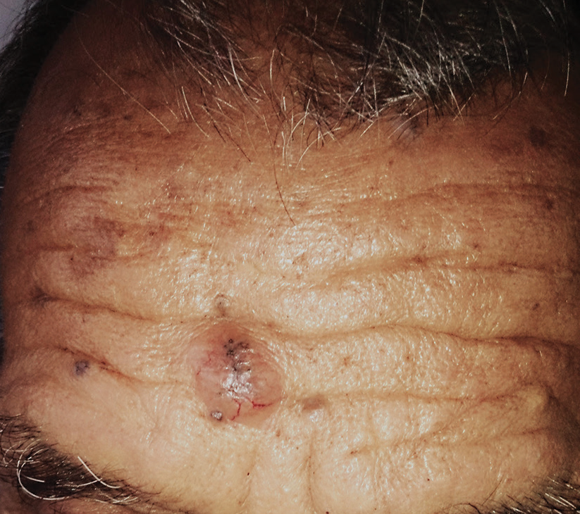
- Well-defined skin colored nodule with telangiectasia on the forehead.
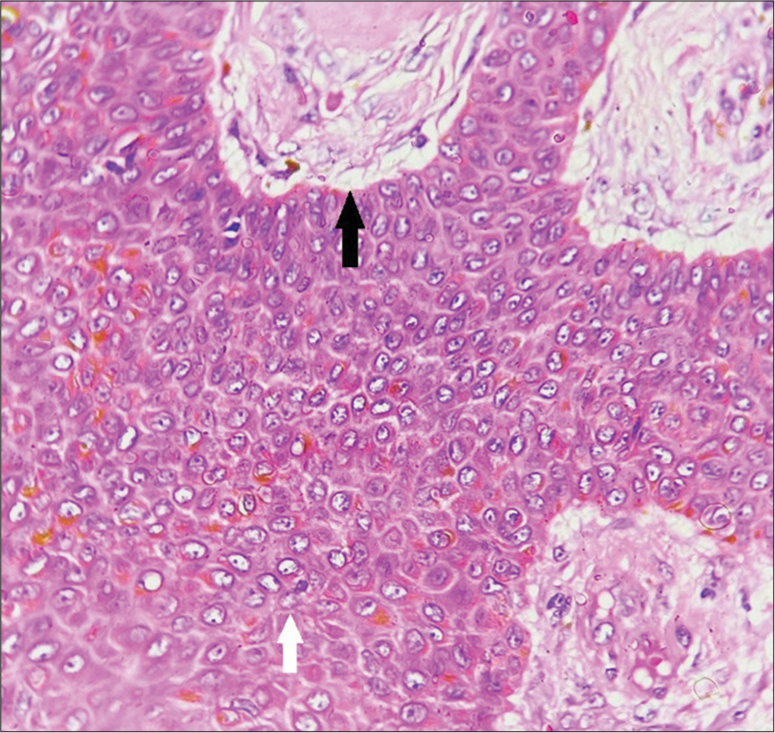
- Small cells with clear cytoplasm and tiny basophilic nuclei (white arrow) and tubular lumina filled with colloid material (black arrow), H&E ×400.
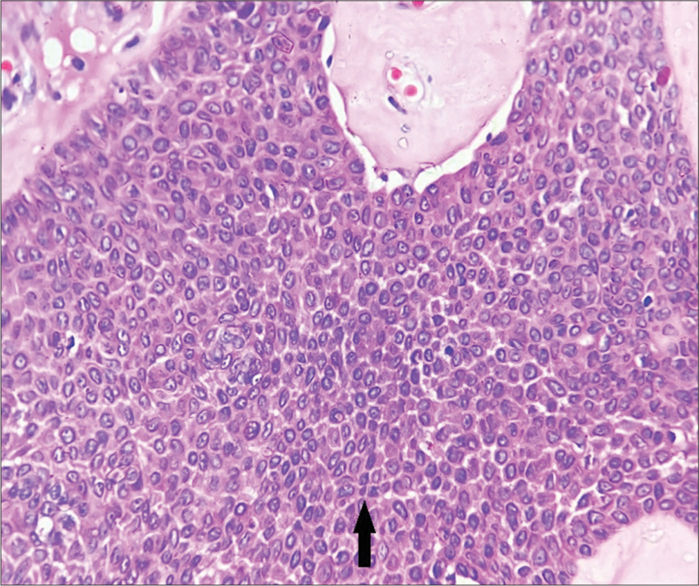
- Fusiform cells with basophilic cytoplasm (arrow), H&E ×400.
Ethical approval
The Institutional Review Board approval is not required.
Declaration of patient consent
The authors certify that they have obtained all appropriate patient consent.
Conflicts of interest
There are no conflicts of interest.
Use of artificial intelligence (AI)-assisted technology for manuscript preparation
The authors confirm that there was no use of artificial intelligence (AI)-assisted technology for assisting in the writing or editing of the manuscript and no images were manipulated using AI.
Financial support and sponsorship
Nil.
References
- An unsual presentation of a nodular hidradenoma. A case report and review of the literature. Ann Med Surg (Lond). 2021;61:61-3.
- [CrossRef] [PubMed] [Google Scholar]


