
Translate this page into:
Papillon–Lefevre syndrome in two brothers: Innocuous or dangerous?


*Corresponding author: Saritha Mohanan, Department of Dermatology, Venereology and Leprosy, Indira Gandhi Medical College and Research Institute, Puducherry, India. saritha_mohanan@yahoo.co.in
-
Received: ,
Accepted: ,
How to cite this article: Sethumadhavan S, Mohanan S, Venkatesan S, Shanmugham D. Papillon–Lefèvre syndrome in two brothers: Innocuous or dangerous? CosmoDerma. 2025;5:46. doi: 10.25259/CSDM_37_2025
Abstract
Two brothers, born of second-degree consanguineous marriage, presented with syndromic diffuse palmoplantar keratoderma with transgrediens and progrediens, along with generalized severe periodontitis, premature loss of permanent teeth and psoriasiform plaques over the elbows, knees, and gluteal region. There was a history of palmoplantar hyperhidrosis and recurrent skin infections. Based on the clinical picture, a diagnosis of Papillon– Lefevre syndrome was made. Pyogenic liver abscesses, cerebral and renal abscesses, and dural calcification are associated complications, which were not present in our patients. Early diagnosis and multidisciplinary approach (systemic retinoids, meticulous dental care, and dentures) are imperative to improve the prognosis.
Keywords
Palmoplantar keratoderma
Papillon–Lefevre syndrome
Periodontitis
Premature loss of teeth
Pyogenic liver abscess

INTRODUCTION
Papillon–Lefevre syndrome is a rare genodermatosis characterized by palmoplantar keratoderma (PPK) and periodontitis. It was initially reported by Papillon and Lefevre in 1924 as a brother and sister.[1] It is an autosomal recessive disorder due to a CTSC mutation on chromosome 11q14, which encodes for cathepsin-C, a lysosomal protease.[2] Parental consanguinity is a contributing factor. More than 300 cases have been reported in literature, with more prevalence among Arabs, Indians, and Africans.[3] Among patients, 20–25% have increased susceptibility to infections, besides periodontitis. It may be associated with malodorous hyperhidrosis, recurrent pyoderma, and nail dystrophy as in our patients.[3] Common complications associated are pyogenic hepatic abscess and calcifications of the dura, falx cerebri, tentorium cerebelli, and choroid plexus of the lateral ventricle.[2,4] Other rare complications include renal abscess and cerebral abscess. We report here two brothers with Papillon–Lefèvre Syndrome (PLS), to highlight the dangerous complications associated with PPK and periodontitis.
CASE REPORT
Case 1
A 19-year-old male, born of second-degree consanguineous marriage, presented with complaints of thickening of palms and soles since 5 years of age, which gradually progressed to involve both knees, elbows, and buttocks. There was also spontaneous gradual loosening and premature loss of most permanent teeth. He also had recurrent pyoderma and palmoplantar hyperhidrosis. He had improvement of PPK when he took isotretinoin for 4 years but recurred on stopping treatment. General examination revealed birdlike facies. Cutaneous examination revealed diffuse thickening of bilateral palms and soles, with fissures, extending to the dorsum of both hands and feet, wrists, and ankles. Symmetrical, well-demarcated, erythematous, and scaly (psoriasiform) plaques were present on both elbows and knees and sparsely over the gluteal region. Nail dystrophy was present in all toenails. Hair and systemic examination were normal. Dental examination revealed loss of almost all permanent teeth and chronic aggressive generalized periodontitis [Figure 1].
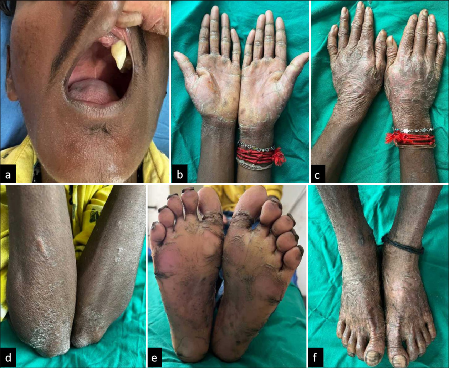
- (a) older brother with loss of most permanent teeth and severe generalized periodontitis, (b and c) diffuse palmoplantar keratoderma extending beyond palms, (d) psoriasiform plaques over elbows and (e and f) soles.
Case 2
A 16-year-old male, younger brother, presented with similar complaints of thickening of palms and soles since 3 years of age, which gradually progressed to involve both knees, elbows, and buttocks, with winter exacerbations. He also had spontaneous gradual loosening and premature loss of many permanent teeth. He also had recurrent pyoderma and palmoplantar hyperhidrosis. He had improvement in skin lesions after isotretinoin intake for 4 years. Cutaneous examination revealed thickening and scaling of bilateral palms and soles, with extensive psoriasiform plaques on the gluteal region. A dental examination revealed multiple missing permanent teeth and chronic aggressive generalized periodontitis, which was less severe than his older brother [Figure 2].

- Younger brother with (a and b) diffuse palmoplantar keratoderma with psoriasiform plaques over (c) elbows and (d) gluteal region and (e) less severe periodontitis and loss of many permanent teeth.
Routine blood investigations such as complete blood count, random blood sugar, renal and liver function tests, and fasting lipid profile were normal. Orthopantomograms in both brothers revealed chronic generalized periodontitis (alveolar bone loss and floating-in-the-air appearance of the teeth). Skin biopsies of the soles of both brothers showed mild hyperkeratosis (orthokeratosis), hypogranulosis, mild regular acanthosis, and sparse perivascular lymphocytic infiltrate in the upper dermis, which was suggestive of nonepidermolytic PPK. Skull X-rays and X-rays of hands and feet were normal [Figure 3]. Ultrasound abdomen was also normal in both brothers.

- Orthopantomogram of younger brother with (a) floating in air appearance, (b) normal X-rays of skull, and (c and d) hands and feet of older brother and biopsy of older brother showing nonepidermolytic palmoplantar keratoderma, hematoxylin and eosin (e) (H&E), ×40, (f) H&E, ×10.
Based on history and clinical examination, a diagnosis of Papillon–Lefevre syndrome was made, and they were started on moisturizers (urea, white soft paraffin, and liquid paraffin), keratolytic (salicylic acid), and antibiotics (whenever they had a secondary infection). Isotretinoin was not restarted due to non-affordability. They were referred to a periodontist for dental hygiene, regular plaque removal (scaling), elective extraction of affected teeth and mandibular and maxillary dentures, and other management. Due to financial constraints, genetic testing was not done.
DISCUSSION
We report here two cases of syndromic diffuse PPK with transgrediens (extends beyond palms and soles) and progrediens (worsens with age), with periodontitis, namely, PLS. PLS is commonly associated with severe progressive periodontitis, which commences between 1 and 4 years of age and peaks in teenage, culminating in premature loss of deciduous and permanent teeth. Other rare features reported that were not present in our case are transverse grooving and fissuring of nails, pseudo-ainhum, follicular hyperkeratosis, thin sparse hair, hepatosplenomegaly, congenital hydrocele, mental retardation, hepatic abscess, cerebral abscess, lung abscess, renal abscess, recurrent tonsillitis, pneumonia, urinary tract infections, squamous cell carcinoma, and melanoma.[1-3,5-7]
Differential diagnosis includes Haim–Munk syndrome and prepubertal periodontitis, both of which are due to cathepsin-C mutation.[1] The former, which is an allelic variation of PLS, has additional features of pes planus, onychogryphosis, arachnodactyly, acro-osteolysis, and more extensive and severe cutaneous features, but less severe periodontitis.[8] Prepubertal periodontitis has no PPK. Pachyonychia congenita presents with natal teeth, oral leukokeratosis, hyperhidrosis, and nail dystrophy, but there is no premature loss of teeth.
Out of more than 300 PLS patients reported, hepatic abscesses have been reported in 22 cases and renal, lung, and cerebral abscesses have been reported in one case each. Hepatic abscess can occur secondary to bacteremia due to periodontitis and impaired immunity and may be recurrent. In PLS, neutrophil and monocyte chemotaxis and phagocytosis, generation of neutrophil extracellular traps, lymphocyte proliferation, reactivity to B- and T-cell mitogens, and cytotoxicity of natural killer cells are impaired.[3,4,7] Common organisms implicated are Staphylococcus aureus and Escherichia coli, with one rare case report of Rhizopus oryzae.[2,4,9] Hepatic abscesses can be treated by drainage and parenteral antibiotics.[9]
Oral retinoids reduce hyperkeratosis and periodontal complications and prevent autoamputation. Keratoderma will persist throughout life. Despite meticulous dental care, all patients eventually become edentulous by 15 years of age.[3] Timely periodontal intervention will prevent early loss of teeth, which, in turn, will prevent jaw bone loss and bird facies. Early identification, systemic retinoids, and a multidisciplinary approach with psychological counseling, dental, and dermatological management can improve the prognosis.[1] Genetic counseling also should be done. These cases are of clinical relevance to dermatologists and physicians because pyogenic hepatic abscesses and other abscesses such as cerebral and renal should be suspected while evaluating pyrexia of unknown origin in PLS.[4]
CONCLUSION
Papillon-Lefevre syndrome can be associated with abscesses of the liver, cerebrum, and kidney. A high index of suspicion is required to detect these complications of this otherwise innocuous syndrome.
Ethical approval
The Institutional Review Board approval is not required.
Declaration of patient consent
The authors certify that they have obtained all appropriate patient consent.
Conflicts of interest
There are no conflicts of interest.
Use of artificial intelligence (AI)-assisted technology for manuscript preparation
The authors confirm that there was no use of artificial intelligence (AI)-assisted technology for assisting in the writing or editing of the manuscript and no images were manipulated using AI.
Financial support and sponsorship: Nil.
References
- Papillon-Lefèvre syndrome: A rare case report of two brothers and review of the literature. Int J Clin Pediatr Dent. 2018;11:352-5.
- [CrossRef] [PubMed] [Google Scholar]
- Diagnosis of Papillon-Lefèvre syndrome: Review of the literature and a case report. Postepy Dermatol Alergol. 2020;37:671-6.
- [CrossRef] [PubMed] [Google Scholar]
- Papillon-Lefèvre syndrome: From then until now. Stomatol Dis Sci. 2019;3:1-16.
- [CrossRef] [Google Scholar]
- Pyogenic liver abscess and Papillon-Lefevre syndrome: Not a rare association. Pediatrics. 2003;111:e85-8.
- [CrossRef] [PubMed] [Google Scholar]
- Papillon-Lefevre syndrome in two brothers. Indian J Dermatol Venereol Leprol. 2002;68:155-6.
- [Google Scholar]
- Multiple cerebral abscesses in Papillon-Lefèvre syndrome. Childs Nerv Syst. 2013;29:1227-9.
- [CrossRef] [PubMed] [Google Scholar]
- Is etretinate dangerous in Papillon-Lefèvre syndrome? Dermatologica. 1988;176:148-50.
- [CrossRef] [PubMed] [Google Scholar]
- Dermatologic, periodontal, and skeletal manifestations of Haim-Munk syndrome in two siblings. J Am Acad Dermatol. 2008;58:339-44.
- [CrossRef] [PubMed] [Google Scholar]
- Pyogenic liver abscess and peritonitis due to Rhizopus oryzae in a child with PapillonLefevre syndrome. Eur J Pediatr. 2011;170:803-5.
- [CrossRef] [PubMed] [Google Scholar]


