
Translate this page into:
Neglected tropical disease presenting as erythroderma in an immunocompetent host – A chronicle of imbalance in Th1/Th2 immune response

*Corresponding author: Yamini Sihag, Department of Dermatology, All India Institute of Medical Sciences, Rajkot, Gujarat, India. yaminisihag174@gmail.com
-
Received: ,
Accepted: ,
How to cite this article: Tyagi S, Sihag Y, Singh A. Neglected tropical disease presenting as erythroderma in an immunocompetent host – A chronicle of imbalance in Th1/Th2 immune response. CosmoDerma. 2024;4:110. doi: 10.25259/CSDM_126_2024
A 60-year-old male without any known comorbidities presented with intense pruritus (Visual Analog Scale 9/10) and redness all over the body for the past 1–1.5 years. It started as nocturnal pruritus over the web spaces of fingers, cubital fossa, axilla, abdomen, and groin and later progressed to involve the entire body with sparing of the face [Figures 1a, b, and c]. Similar complaints were also present in his family members and peers. Based on the above presentation, we suspected the case to be chronic erythroderma secondary to Norwegian scabies. A bedside test of potassium hydroxide mount of skin scrapings showed Sarcoptes scabiei mite thus confirming the diagnosis [Figure 1d]. Given the association of crusted/Norwegian scabies with immunosuppression, the patient was evaluated but no conclusive evidence was found on clinical and laboratory examination. The patient received treatment as per Centers for Disease Control and Prevention guidelines for Norwegian scabies, that is, cream permethrin 5% (daily application for 7 days, then 2 times/week till cure) with oral ivermectin (200 microgram/kg body weight on day 1, 2, 8, 9, and 15) and antihistamines and showed dramatic improvement in 2 weeks of treatment.
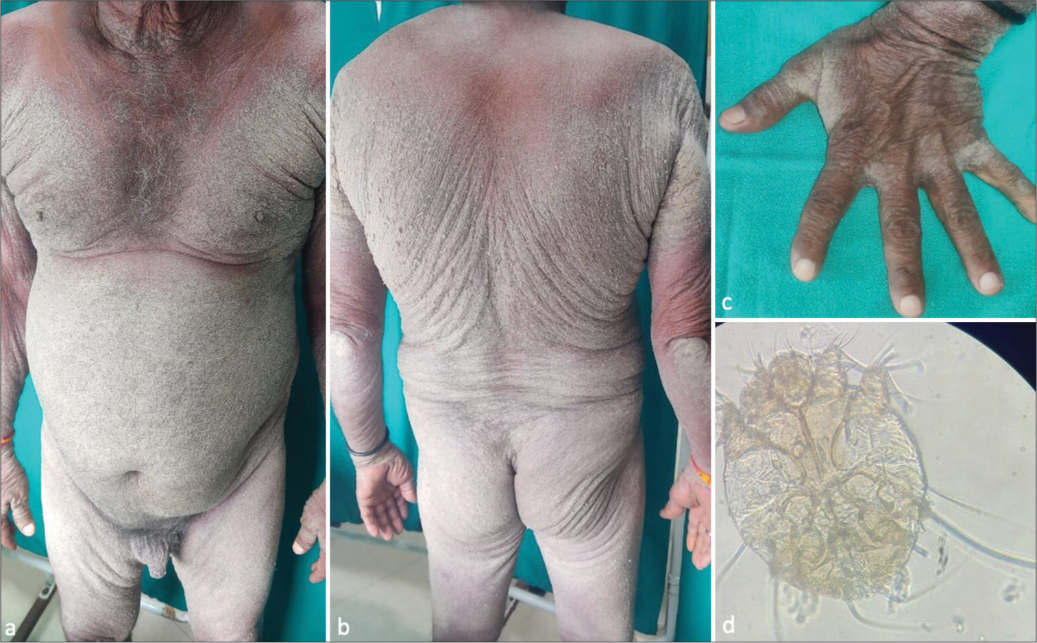
- (a) Diffuse erythema with thick yellowish-white crust over the abdomen, genitals, and upper limbs. (b) Diffuse erythema with thick yellowish-white crust over the back, gluteal area, upper limbs, and lower limbs. (c) Finger webs showing yellowish-white crust. (d) Potassium hydroxide mount of skin scrapings showing Sarcoptes scabiei mite (visualized under ×10 magnification using Olympus CX21i microscope).
Erythroderma refers to diffuse erythema and scaling of skin involving >90% of body surface area. It occurs commonly secondary to psoriasis (19%), drug reactions (19%), atopic dermatitis (9%), cutaneous T-cell lymphoma (8%), contact dermatitis (6%), seborrheic dermatitis (4%), and less commonly (<0.5%) due to scabies, lichen planus, and dermatomyositis.[1,2] Norwegian scabies or crusted scabies is a fulminant infestation of the female gravid mite of S. scabiei. presenting as thick crusted plaques, fissures with or without secondary bacterial infection due to the absence of adequate immune response. It is generally seen in cases of immunosuppression, sensory anesthesia, mental or physical impairment, treated leprosy, substance abuse, systemic lupus erythematosus, pulmonary Koch’s disease, diabetes mellitus, and hepatitis B.[2,3] It is rare for immunocompetent individuals to develop crusted scabies. Ordinary scabies has a Th1 immune response while crusted scabies is known to elaborate a Th2 immune response. The CD4+ T-cells of the Th1 immune response which secretes interferon-γ and interleukin (IL)-2 can redirect the Th2 immune response, that is, CD4+ T-cells secreting IL-4, IL-5, IL-6, and IL-13. This concept has been extended to CD8+ T-cells as well, which are predominant cells seen in crusted scabies giving rise to a higher ratio of CD8+ to CD4+ T-cells. Activated CD8+ T-cells in crusted scabies induce disrupted keratinocyte apoptosis leading to hyperkeratotic lesions. These cells also secrete cytokines to target resident skin cells and amplify inflammation. Increased transforming growth factor-beta expression in crusted scabies is also responsible for suppressing Th1 responses. Therefore, crusted scabies develops secondary to an imbalance in the Th1/Th2 immune response.[3] In the absence of immunocompromised states, diagnosis of crusted scabies may be missed in an otherwise immunocompetent patient. The present case manifests this scarcely reported phenomenon of ordinary scabies transforming into Norwegian scabies and presenting as chronic erythroderma in an immunocompetent male.
Ethical approval
The Institutional Review Board approval is not required.
Declaration of patient consent
The authors certify that they have obtained all appropriate patient consent.
Conflicts of interest
There are no conflicts of interest.
Use of artificial intelligence (AI)-assisted technology for manuscript preparation
The authors confirm that there was no use of artificial intelligence (AI)-assisted technology for assisting in the writing or editing of the manuscript and no images were manipulated using AI.
Financial support and sponsorship
Nil.
References
- Erythrodermic scabies in an immunocompetent patient. JAAD Case Rep. 2022;29:112-5.
- [CrossRef] [PubMed] [Google Scholar]
- Norwegian scabies: Rare cause of erythroderma. Indian Dermatol Online J. 2015;6:52-4.
- [CrossRef] [PubMed] [Google Scholar]
- New insights into disease pathogenesis in crusted (Norwegian) scabies: The skin immune response in crusted scabies. Br J Dermatol. 2008;158:1247-55.
- [CrossRef] [PubMed] [Google Scholar]


