
Translate this page into:
Kerion celsi caused by Trichophyton mentagrophytes: Clinical, trichoscopic, and microscopic features

*Corresponding author: Vishal Gaurav, Department of Dermatology and Venereology, Maulana Azad Medical College, New Delhi, India. mevishalgaurav@gmail.com
-
Received: ,
Accepted: ,
How to cite this article: Lalmuanpuii G, Barman KD, Das S, Gaurav V. Kerion celsi caused by Trichophyton mentagrophytes: Clinical, trichoscopic, and microscopic features. CosmoDerma. 2024;4:70. doi: 10.25259/CSDM_69_2024
A 6-year-old boy presented with a single painful boggy swelling with overlying hair loss on the scalp for two weeks. On examination, there was a single, well-defined, erythematous , fluctuant nodular swelling over the mid-frontal scalp measuring 4 × 4 cm, with multiple follicular pustules, semi-adherent yellowish-white crusts, peripheral white scales, and overlying alopecia [Figure 1] without any regional lymphadenopathy. Trichoscopy revealed multiple yellow globules, few pigtail, corkscrew as well as morse code hair, and black dots along with hemorrhage [Figure 2], over a background of erythema. Microscopy of potassium hydroxide (KOH) mount of a plucked hair revealed intrapilary arthroconidia [Figure 3]. Trichophyton mentagrophytes complex was isolated on culture in Sabouraud dextrose agar at 25° C. Lactophenol cotton blue mount from the culture showed spiral hyphae [Figure 4]. Based on the clinical, trichoscopic, and microscopy features, a diagnosis of kerion caused by T. mentagrophytes was made and the patient was started on oral griseofulvin microsize suspension at a dose of 20 mg/kg for 6 weeks. However, the response to therapy could not be assessed as the patient was lost to follow-up.

- Single, well-defined, erythematous nodular swelling over the mid-frontal scalp measuring 4 × 4 cm, with multiple follicular pustules, semi-adherent yellowish-white crusts, peripheral white scales, and overlying alopecia.
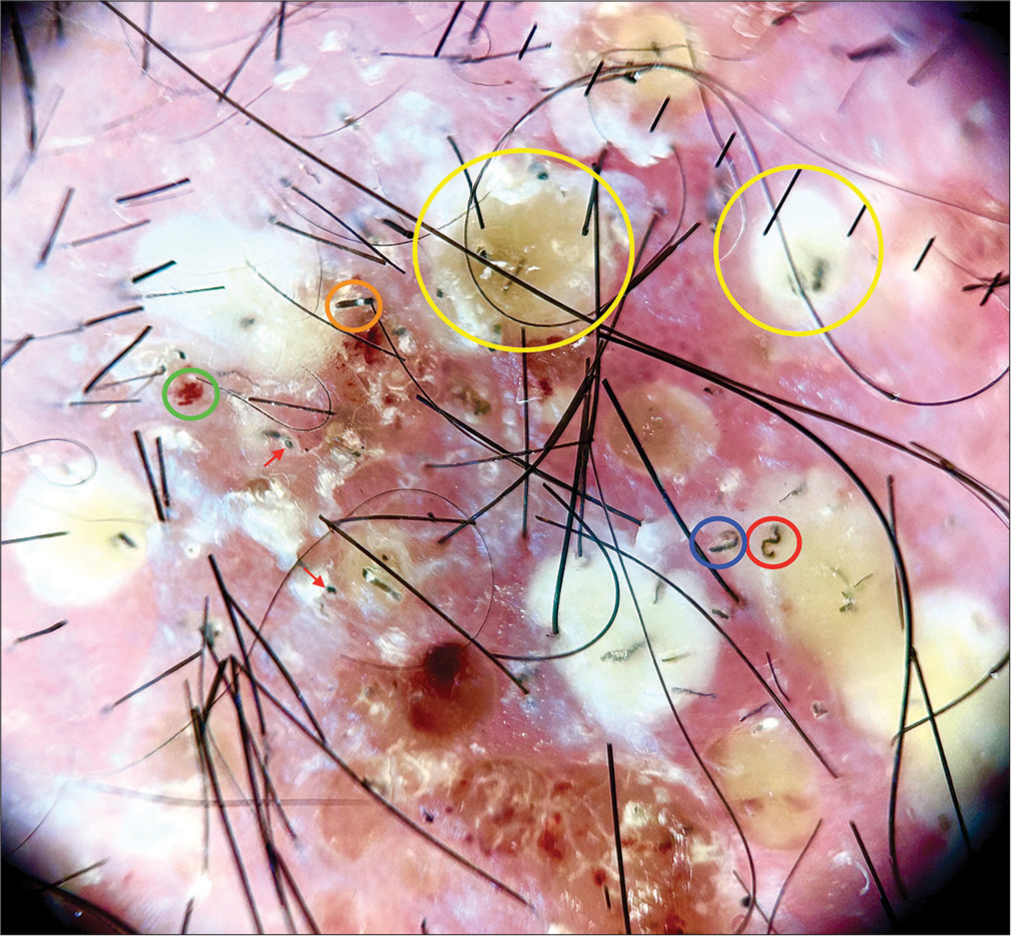
- Trichoscopic examination in polarized mode (Heine DELTAone) at ×10 magnification showing multiple yellow globules (yellow circles), few pigtail (navy blue circle), corkscrew (red circle) as well as morse code hair (orange circle), and black dots (red arrows) along with hemorrhage (green circle), over a background of erythema.
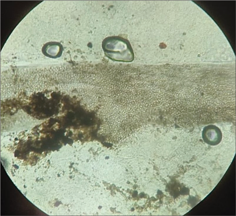
- Microscopy of potassium hydroxide mount of a plucked hair showing intrapilary arthroconidia (×400).
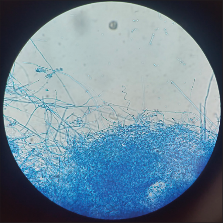
- Microscopy of lactophenol cotton blue mount from the culture on Sabouraud dextrose agar at 25° C showing spiral hyphae (×100).
Kerion, an inflammatory tinea capitis, presents as boggy, tender nodules with pustules, scaling, alopecia, and regional lymphadenopathy, mostly in children. Common causative agents include Trichophyton tonsurans and Microsporum canis, with T. mentagrophytes being uncommon.[1] Diagnosis involves dermoscopy showing yellow dots, comma hairs, and corkscrew hairs, and KOH microscopy revealing arthroconidia and septate hyphae.[2] Treatment with oral antifungals (griseofulvin, terbinafine, and itraconazole) is essential to prevent scarring alopecia. Prognosis is generally good with timely intervention.[1]
Ethical approval
The Institutional Review Board approval is not required.
Declaration of patient consent
The authors certify that they have obtained all appropriate patient consent.
Conflict of interest
There are no conflicts of interest.
Use of artificial intelligence (AI)-assisted technology for manuscript preparation
The authors confirm that there was no use of artificial intelligence (AI)-assisted technology for assisting in the writing or editing of the manuscript and no images were manipulated using AI.
Financial support and sponsorship
Nil.
References
- Kerion Celsi in infants and children-A narrative review 2010-2023. Mycoses. 2024;67:e13675.
- [CrossRef] [PubMed] [Google Scholar]
- The dermatoscope in the hair clinic: Trichoscopy of scarring and nonscarring alopecia. J Am Acad Dermatol. 2023;89:S9-15.
- [CrossRef] [PubMed] [Google Scholar]


