
Translate this page into:
Kerion caused by Microsporum species – Diagnosed by Wood’s lamp and microscopy

*Corresponding author: Pradeep S. Nair, Department of Dermatology and Venereology, Government TD Medical College, Alappuzha, Kerala, India. dvmchtvm@yahoo.co.in
-
Received: ,
Accepted: ,
How to cite this article: Nair PS. Kerion caused by Microsporum species – Diagnosed by Wood’s lamp and microscopy. CosmoDerma. 2024;4:127. doi: 10.25259/CSDM_148_2024
A 7-year-old boy presented with a swelling on the scalp of a 3-week duration. Initially, the boy had itching on the central part of the scalp followed by pain, pustulation, and crusting. Within a period of two weeks, the lesion gradually progressed to attain the present size. There was no history of trauma or contact with pets. On examination, there was a well-defined tender boggy, soft to firm swelling about 5 × 4 cm on the central part of the scalp with the surface studded with pustules, crusting, and loss of hair [Figure 1]. The hairs at the periphery of the boggy mass were broken with crusts on the shaft. There was cervical lymphadenopathy. Microscopic examination of the hair at the periphery showed the hair shaft studded with small spores suggestive of ectothrix [Figure 2a]. Wood’s lamp examination demonstrated bright green fluorescence of the boggy mass suggestive of Microsporum species [Figure 2b]. Fungal culture was positive for Microsporum canis species. We made a final diagnosis of kerion caused by M. canis. The boy was treated with systemic fluconazole, terbinafine cream, and ketoconazole shampoo. Kerion is a severe inflammatory tinea capitis caused by zoophilic and geophilic species of dermatophytosis. The usual species are M. canis (most common), Microsporum audouinii, Trichophyton verrucosum, and Trichophyton mentagrophytes.[1] Here, we could diagnose the generic species by simple microscopic examination (ectothrix) and Wood’s lamp (bright green fluorescence), features of Microsporum, confirmed by culture, while Trichophyton species caused endothrix and some species cause pale yellow fluorescence on Wood’s lamp examination.
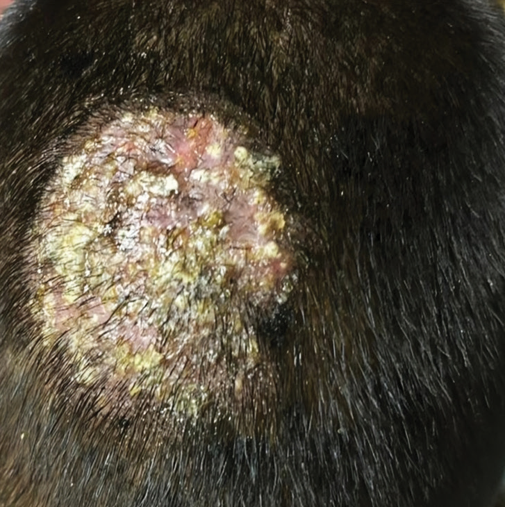
- Well-defined boggy swelling studded with pustules and crusting on central scalp.
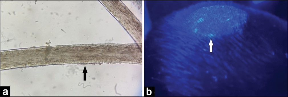
- (a) Hair microscopy showing spores on the shaft (arrow) suggestive of ectothrix, potassium hydroxide ×100, (b) Wood’s lamp examination showing bright green fluorescence suggestive of Microsporum species (arrow).
Ethical approval
Institutional Review Board approval is not required.
Declaration of patient consent
The authors certify that they have obtained all appropriate patient consent.
Conflicts of interest
There are no conflicts of interest.
Use of artificial intelligence (AI)-assisted technology for manuscript preparation
The authors confirm that there was no use of artificial intelligence (AI)-assisted technology for assisting in the writing or editing of the manuscript and no images were manipulated using AI.
Financial support and sponsorship
Nil.
References
- Kerion celsi in a Nepalese boy: An underdiagnosed cause of scalp swelling. Case Rep Infect Dis. 2021;24:59-63.
- [CrossRef] [Google Scholar]


