
Translate this page into:
Diffuse loss of hair with papules

*Corresponding author: Gajanand M. Antakanavar, Department of Dermatology, Maulana Azad Medical College, Delhi, India. gajanand.hooli@gmail.com
-
Received: ,
Accepted: ,
How to cite this article: Antakanavar GM, Relhan V, Chauhan N. Diffuse loss of hair with papules. CosmoDerma. 2024;4:116. doi: 10.25259/CSDM_143_2024
A 20-year-old male, born to non-consanguineous parents, presented with complete loss of hair on the scalp and body since the age of two months. In addition, at three years old, he developed skin-colored papules on his face and body. These papules initially appeared on the scalp and gradually progressed to involve the rest of the body within a span of two years. These lesions were unresponsive to treatment. There was no history suggestive of delay in milestones, decreased sweating, diminution of vision, decreased hearing, seizures, atopy, or bone pains. There was no family history of a similar illness. Clinical examination revealed a complete absence of hair on the scalp and entire body including the axilla and groin [Figure 1a-c]. Multiple discrete skin-colored smooth and milia-like papules of size ranging from 0.5 cm to 1 cm distributed on the whole body. Dermatoscopy of the scalp and forearm showed yellow dots, an absence of hair follicle ostia, and a cyst-like lesion, which is giving a cluster of star appearance [Figure 1d]. Mucosa, nails, teeth, palms, and soles were normal. There were no bony abnormalities, dysmorphic features, or any systemic involvement. Based on clinical history and examination, differential diagnoses of atrichia with papular lesion (APL) and alopecia areata universalis were made. Serum Vitamin D, calcium, phosphate, and parathormone levels were within normal limits. Histopathological examination of scalp biopsy [Figure 1e and f] unveiled multiple dermal cysts containing keratinous material, absence of normal hair follicles, and signs of inflammation.
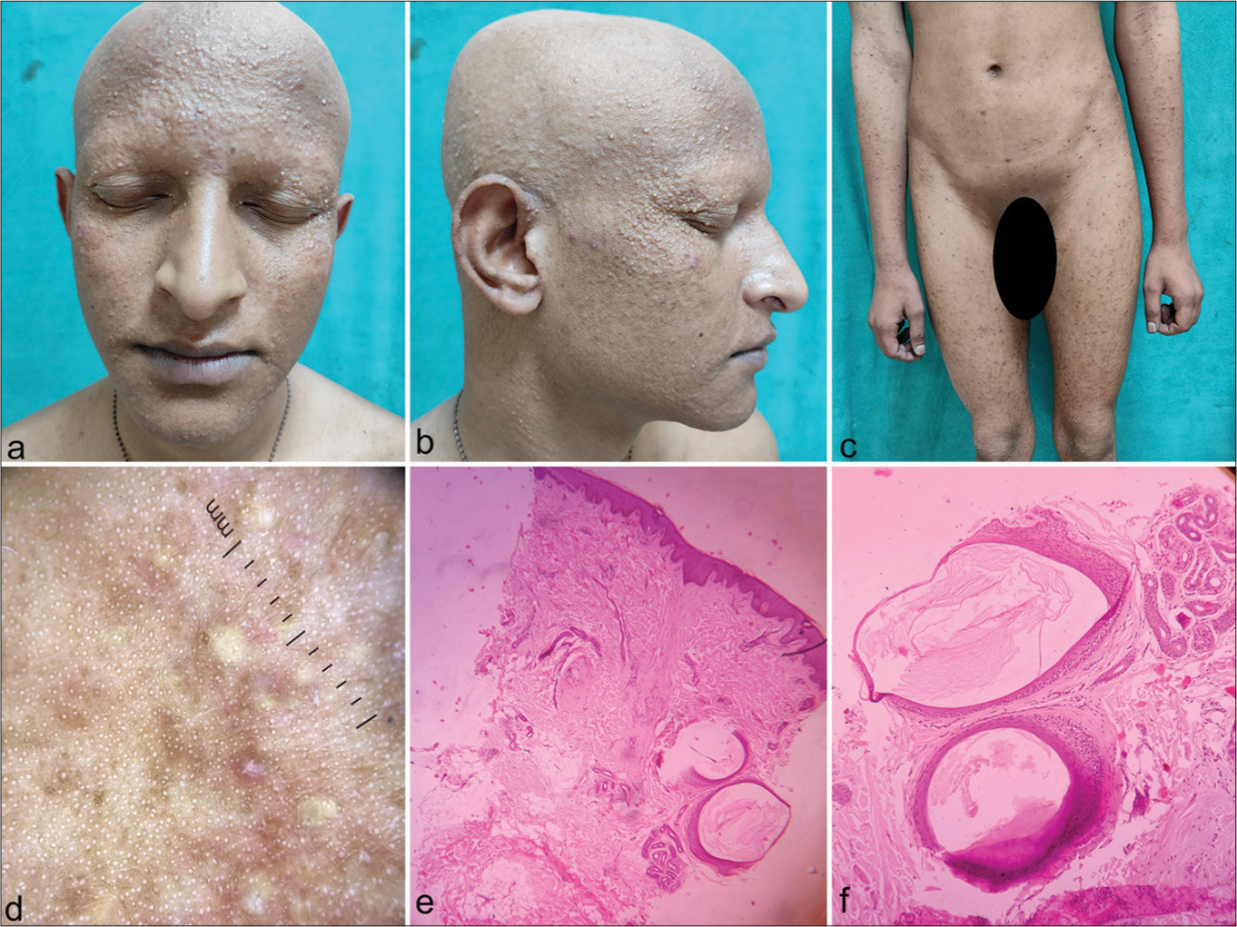
- (a) Complete absence of hair of scalp, eyebrows, eyelashes, beard, and moustache. (b) Multiple tiny milia like discrete papules were distributed over scalp, lateral canthus along with complete loss of hair and (c) similar papules present over abdomen and thigh along with absence of pubic hair. (d) Dermatoscopy revealed cluster of star appearance along with loss of follicular ostia. (e) Histopathological examination of biopsy from scalp revealed (H&E staining, ×100), complete absence of normal hair follicles, and absence of signs of inflammation. Dermis showed dermal cysts containing keratinous material. (f) Magnified view of dermis (×400) revealed dermal cysts lined by stratified squamous epithelium, which shows keratinous material inside.
WHAT IS THE DIAGNOSIS?
Answer:
Atrichia with papular lesions
DISCUSSION
Atrichia with papular lesions is a rare autosomal recessive follicular disorder caused by non-sense mutations of the hairless (HR) gene.[1] However, heterozygous individuals have normal hair and are clinically indistinguishable from genotypically normal persons. In APL, along with HR gene mutation, there occurs a decline in B-cell lymphoma expression and loss of neural cell adhesion molecule positivity. This results in hair matrix cells proximal inner and outer root sheath undergoing a premature and massive apoptosis and disconnection from the overlying epithelial sheath which is essential for the movement of the dermal papilla.[2] As a consequence, the hair bulbs and dermal papillae remain stranded in the dermis, and signals are not transferred between dermal papillae and stem cells in the bulge. This halts the further growth of hair follicles. Hence, hair shafts are not formed at the end of the anagen phase.[2]
Lanugo hairs are initially present at birth, but alopecia becomes complete within the first year of life as catagen follicles are unable to re-enter the anagen phase. Progressive keratin retention into follicles results in cystic formations clinically evident as papules.[1] The rarer conditions such as alopecia universalis (AU) congenita and Hereditary Vitamin D–Resistant Rickets (HVDRR) can mimic clinically and histologically APL. However, HVDRR is usually asymptomatic at birth and during the first 2 years of life, which displays delayed neuromotor development, bone abnormalities, and frequent infections.[3] Hair loss may precede the development of bony rachitic changes.[4] The index case met four out of five major criteria as per Yip et al.[1] [Table 1]. However, genetic testing for the HR gene and Vitamin D resistance could not be done due to inadequate facilities. The development of AU within the first year of life, disseminated cysts or papules, in the absence of rachitic changes, and non-responsiveness to therapy all together strongly suggest the diagnosis of APL. Atrichia with papular lesion mimics an autoimmune form of AU. Most cases are often incorrectly treated with steroids or immunosuppressants. When dealing with cases of universal alopecia with papules that are resistant to therapy, it becomes imperative to consider APL. Therefore, careful clinical, dermatoscopy, and histopathological examination will guide in diagnosing such rare cases.
| Major Criteria (4 out of 5 required for diagnosis) |
| 1) Permanent and complete absence of scalp hairs by the first few months of life. |
| 2) Few to widespread smooth, whitish, or milia-like papules on the face, scalp, arms, elbows, thighs or knees from infancy or childhood. |
| 3) Replacement of mature hair follicle structures by follicular cysts filled with cornified material in scalp histology. |
| 4) Mutation(s) in the human hairless gene through genetic testing |
| 5) Clinical and/or molecular exclusion of vitamin-D-dependent rickets |
| Minor Criteria (Supplementary criteria) |
| 1) Family history of consanguinity |
| 2) Absence of secondary axillary, pubic, or body hair growth and/or sparse eyebrows and eyelashes. |
| 3) Normal growth and development, including normal bones, teeth, nails and sweating. |
| 4) Whitish-hypopigmented streaks on the scalp. |
| 5) Lack of response to any treatment modality |
Ethical approval
The Institutional Review Board approval is not required.
Declaration of patient consent
The authors certify that they have obtained all appropriate patient consent.
Conflicts of interest
There are no conflicts of interest.
Use of artificial intelligence (AI)-assisted technology for manuscript preparation
The authors confirm that there was no use of artificial intelligence (AI)-assisted technology for assisting in the writing or editing of the manuscript and no images were manipulated using AI.
Financial support and sponsorship
Nil.
References
- Atrichia with papular lesions: A report of three novel human hairless gene mutations and a revision of diagnostic criteria. Acta Derm Venereol. 2008;88:346-9.
- [CrossRef] [Google Scholar]
- Atrichia congenita with papular lesions. Indian J Dermatol Venereol Leprol. 2011;77:70.
- [CrossRef] [Google Scholar]
- Alopecia in patients with vitamin D-resistant rickets type-II. An Bras Dermatol. 2017;92:286-7.
- [CrossRef] [Google Scholar]
- Atrichia caused by mutations in the vitamin D receptor gene is a phenocopy of generalized atrichia caused by mutations in the hairless gene. J Invest Dermatol. 2001;117:612-7.
- [CrossRef] [Google Scholar]


