Translate this page into:
Differential mosaicism of hypertrichosis and hypomelanosis

*Corresponding author: Niharika Dhattarwal, Department of Dermatology, Chacha Nehru Bal Chikitsalaya, Delhi, India. niharika.pgirtk@gmail.com
-
Received: ,
Accepted: ,
How to cite this article: Dhattarwal N, Singh B. Differential mosaicism of hypertrichosis and hypomelanosis. CosmoDerma. 2024;4:96. doi: 10.25259/CSDM_80_2024
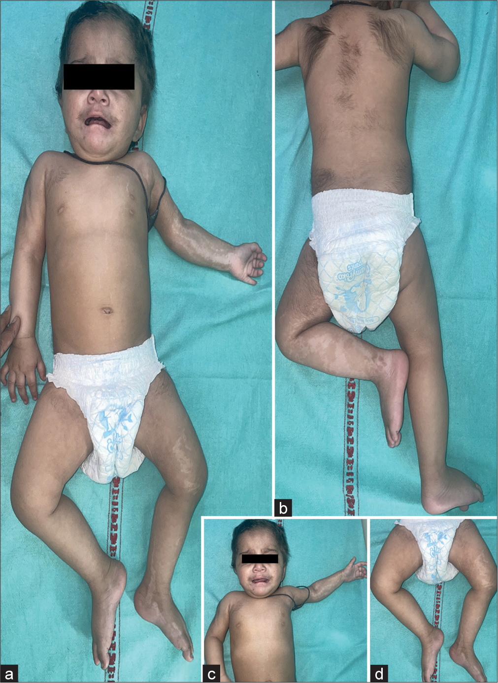
A 1-year-old baby girl presented with increased hair growth and white spots on her body since birth [Figure 1a]. The baby was born out of a consanguineous marriage at 38 weeks. There were no antenatal or intrapartum complications. She has two older siblings, who do not have similar complaints. There were no other cutaneous or systemic complaints. On examination, patchy hypertrichosis was noted over the face, limbs, and trunk [Figure 1b] along with blaschkoid hypopigmentation over the left upper and lower limb [Figure 1c and d]. Systemic examination (central nervous system, cardiovascular system, respiratory, per abdomen, and genitourinary), musculoskeletal (observable anatomic anomalies and active range of motion of joints), ocular and dental examination, anthropometry, and developmental milestone evaluation were within normal limits. Diagnosis of multiple nevoid hypertrichosis was made based on distribution and ruling out other causes of hypertrichosis. Hypomelanosis of Ito was diagnosed due to characteristic blaschkoid hypopigmentation.

- (a) Increased hair growth over face and body and white spots over left side of body, (b) multiple patchy hypertrichosis, (c) Blaschkoid hypopigmentation on the left upper limb, and (d) Blaschkoid hypopigmentation on the left lower limb.
Mosaicism refers to the presence of two or more genetically different sets of cells in an individual. Cutaneous mosaicisms manifest as specific distribution patterns over the skin like blaschkoid, linear, segmental, localized (nevoid), checkerboard, phylloid, and large patches without midline separation and lateralization. Nevoid hypertrichosis is one such entity characterized by a localized patch of terminal hair growth. It is usually present at birth and may be associated with lipoatrophy and bony abnormalities.[1]
Hypomelanosis of Ito is characterized by hypopigmentation along the lines of Blaschko. Extracutaneous findings include neurological, musculoskeletal, dental, and ocular defects.[1]
Twin spotting refers to the co-occurrence of two nevoid skin conditions occurring simultaneously but mutually exclusive of each other, over a background of normal skin. The patient described here is an interesting case of differential mosaicism in two halves of the body. The right side of the body has two genetic populations of cells: One having normal skin and hair and the other having normal skin with hypertrichosis. The left side of the body has three genetically different types of cells: One having normal skin and hair, one having hypopigmented skin with normal hair, and one having normal skin with hypertrichosis. A multidisciplinary approach is warranted for such cases as they may have associated abnormalities. Detailed history, cutaneous examination (including skin, hair, and nails), and clinical examination of all systems, anthropometry and milestones evaluation by a pediatrician is a must. Musculoskeletal, ocular, and dental examinations should also be done. Further, investigations including hematological, biochemical investigations, brain imaging, chest x-ray, ultrasound abdomen, electrocardiogram, x-ray of bones and joints, and pure-tone audiometry are to be done if necessary. Early diagnosis and treatment of associated conditions would improve prognosis.
Ethical approval
The Institutional Review Board approval is not required.
Declaration of patient consent
The authors certify that they have obtained all appropriate patient consent.
Conflicts of interest
There are no conflicts of interest.
Use of artificial intelligence (AI)-assisted technology for manuscript preparation
The authors confirm that there was no use of artificial intelligence (AI)-assisted technology for assisting in the writing or editing of the manuscript and no images were manipulated using AI.
Financial support and sponsorship
Nil.
References
- Hypomelanosis of Ito and multiple naevoid hypertrichosis: Rare cutaneous mosaicism. Australas J Dermatol. 2014;55:e29-32.
- [CrossRef] [PubMed] [Google Scholar]


