
Translate this page into:
Dermoscopy of plantar wart


*Corresponding author: Vishal Gaurav, Department of Dermatology and Venereology, Maulana Azad Medical College, New Delhi, India. mevishalgaurav@gmail.com
-
Received: ,
Accepted: ,
How to cite this article: Gaurav V, Agrawal I. Dermoscopy of plantar wart. CosmoDerma. 2024;4:107. doi: 10.25259/CSDM_104_2024
A 30-year-old female office worker with no significant medical history and no immunosuppression presented with a painful lesion on her left sole for 3 months. The lesion was more painful on walking. Clinical examination revealed a tender hyperkeratotic papule [Figure 1]. The pain was more on lateral pressure compared to vertical pressure. Dermoscopy revealed multiple dotted and linear vessels surrounded by yellow-gray structureless areas giving a “frogspawn appearance” with interrupted and distorted dermatoglyphics over the lesion [Figure 2]. Based on the clinical and dermoscopic findings, a diagnosis of plantar wart was confirmed and the patient was advised to apply a combination of salicylic acid (16.7%) and lactic acid (16.7%) paint once daily.
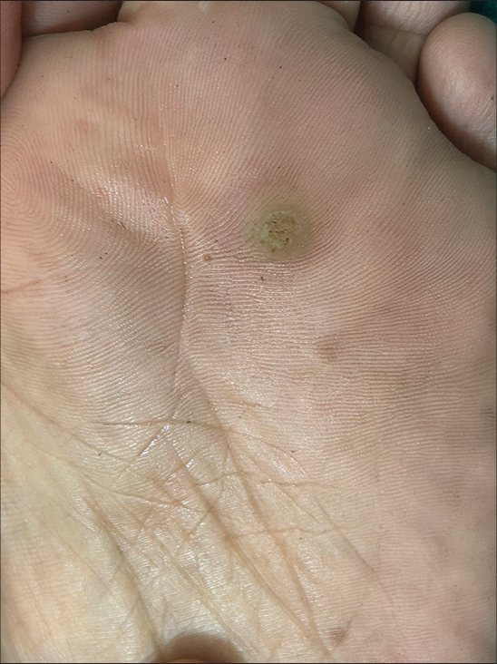
- Solitary hyperkeratotic papule of the forefoot.
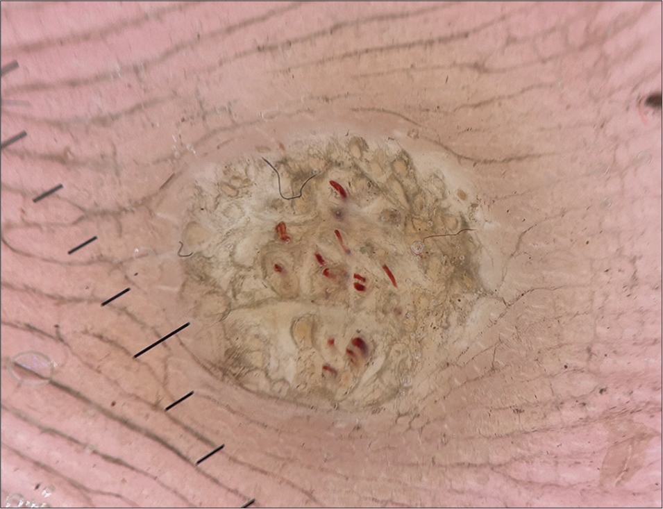
- Polarized dermoscopy (Heine DELTAone) showing multiple dotted and linear vessels surrounded by yellow-gray structureless areas giving a “frogspawn appearance” with interrupted and distorted dermatoglyphics over the lesion (×20).
Plantar warts, or verrucae plantaris, are hyperkeratotic papules caused by HPV, predominantly types 1, 2, 4, and 63. These warts often cause significant pain and can impede walking. Dermoscopy significantly enhances the diagnostic accuracy of plantar warts by revealing specific morphological features not visible to the naked eye. The presence of black dots and globules, dotted and linear vessels, frogspawn or falooda seed appearance, and interrupted dermatoglyphics are highly indicative of warts.[1] These features help differentiate warts from other benign (corns, calluses), and malignant lesions (squamous cell carcinoma). Corns and calluses typically show a central core with intact dermatoglyphics and lack thrombosed capillaries. Squamous cell carcinoma may present with a similar hyperkeratotic appearance but lacks the characteristic black dots and vascular structures of warts.[1] Dermoscopy can be utilized to evaluate the therapeutic response of trichloroacetic acid in treating warts. Successful treatment is indicated by a uniform white frost and the absence of blood vessels on dermoscopic examination, thus improving treatment assessment and efficacy.[2]
Ethical approval
The Institutional Review Board approval is not required.
Declaration of patient consent
The authors certify that they have obtained all appropriate patient consent.
Conflicts of interest
There are no conflicts of interest.
Use of artificial intelligence (AI)-assisted technology for manuscript preparation
The authors confirm that there was no use of artificial intelligence (AI)-assisted technology for assisting in the writing or editing of the manuscript and no images were manipulated using AI.
Financial support and sponsorship
Nil.
References
- Dermoscopy features of cutaneous warts. Int J Gen Med. 2021;14:9903-12.
- [CrossRef] [PubMed] [Google Scholar]
- Utilizing dermoscopy for trichloroacetic acid (TCA) therapy assessment in genital verruca. Indian Dermatol Online J. 2024;15:706-7.
- [CrossRef] [PubMed] [Google Scholar]


