Translate this page into:
Blaschkoid lichen planus

*Corresponding author: Dr. Kurat Sajad, Department of Dermatology, Venereology and Leprosy, Government Medical College, Srinagar, Jammu and Kashmir, India. drkuratsajad@gmail.com
-
Received: ,
Accepted: ,
How to cite this article: Ul Islam M, Sajad K, Farooq M, Saqib N. Blaschkoid lichen planus. CosmoDerma. 2024;4:145. doi: 10.25259/CSDM_172_2024
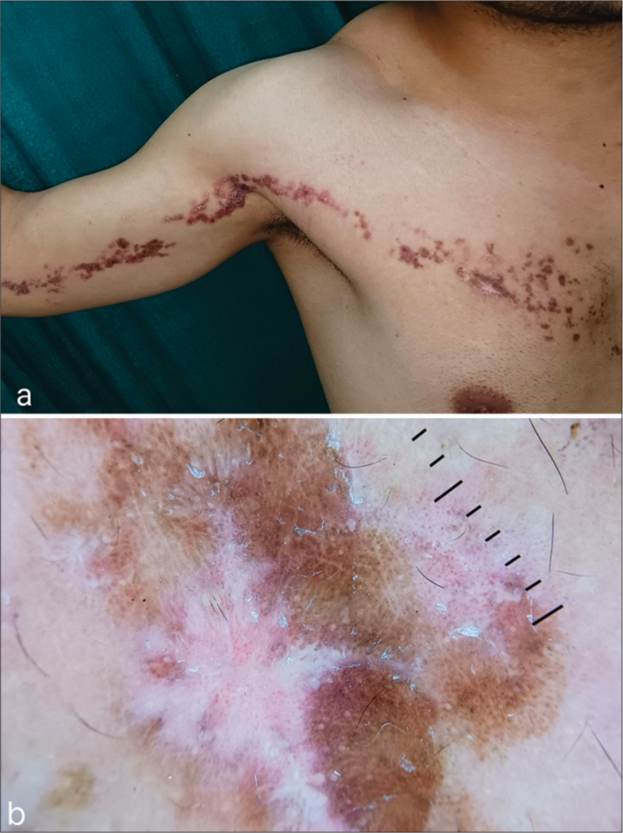
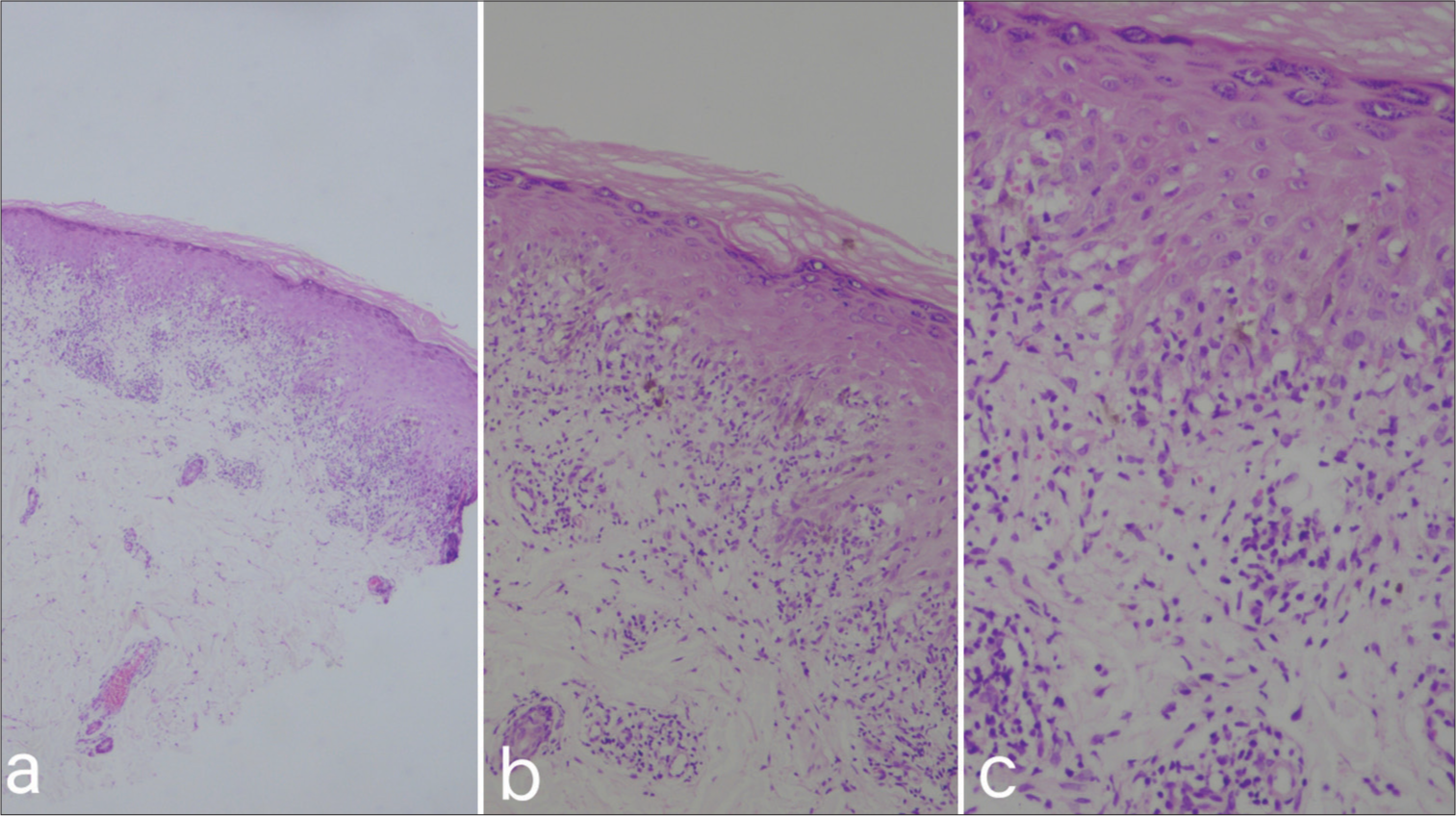
A 28-year-old male student with no comorbidities presented to the dermatology outpatient department with the complaint of pruritic eruptions for the past 4 months over the right arm and extending over the chest. Negative history included no history of travel, recent immunizations, herpes infections, or any significant medication history. On examination, a linear plaque extended from the anterior aspect of the chest, the anterior aspect of the right arm, and the mid-forearm. It was violaceous to brownish with an erythematous background and a few discrete papules in the peripheral surroundings. White scaling was also present over the plaque [Figure 1a]. No oral involvement was present. Dermoscopic examination revealed reticulate whitish structures, white scales, and dotted vessels [Figure 1b]. The patient was subjected to an incisional biopsy, which revealed hyper orthokeratosis, focal hypergranulosis, focal epidermal hyperplasia, vacuolar degeneration of the basal layer, lymphocytic infiltrate at dermo-epidermal junction, and dermal melanophages, which were suggestive of lichen planus (LP) [Figure 2a-c].

- (a) A linear violaceous to brownish plaque with few discrete papules in a blaschkoid pattern extending from the right side of the anterior aspect of the chest to the right arm with an erythematous background and overlying whitish scaling at a few places. (b) Dermoscopic examination showing reticulate whitish structures, white scales and dotted vessels (DL4,10X).

- HPE showing (a and b) hyperorthokeratosis, focal hypergranulosis, focal epidermal hyperplasia, vacuolar degeneration of the basal layer, lymphocytic infiltrate at dermo-epidermal junction, and dermal melanophages (×4, ×10, H&E), (c) lymphocytic infiltrate at dermo-epidermal junction, and dermal melanophages (×40, H&E). H&E: Hematoxylin and eosin, HPE: Histopathological examination..
Blaschkoid LP (syn. linear LP, Blaschko linear LP, and Blaschkoid LP.) is an inflammatory dermatosis comprising only 0.24–0.62% of all cases of LP. It presents as pruritic papules and plaques in a linear pattern following the lines of Blaschko. The suggested mechanism behind this variant is postzygotic mosaic alteration, which leads to a loss of heterozygosity and subsequently leads to the formation of keratinocyte clones, most notorious for developing LP on exposure to triggers. Close differential includes lichen striatus, which predominantly occurs in adolescents. It usually presents as an asymptomatic linear papule/plaque arranged in a blaschkoid pattern with slight scaling mainly involving the proximal part of limbs, and it has a spontaneous resolution over 3–6 months.[1,2]
Ethical approval
Institutional Review Board approval is not required.
Declaration of patient consent
The authors certify that they have obtained all appropriate patient consent.
Conflicts of interest
There are no conflicts of interest.
Use of artificial intelligence (AI)-assisted technology for manuscript preparation
The authors confirm that there was no use of artificial intelligence (AI)-assisted technology for assisting in the writing or editing of the manuscript and no images were manipulated using AI.
Financial support and sponsorship
Nil.
References
- Blaschkoid lichen planus: Throwing a “curve” in the nomenclature of linear lichen planus. JAAD Case Rep. 2020;6:237-9.
- [CrossRef] [PubMed] [Google Scholar]
- Unilateral blaschkoid lichen planus. Indian Dermatol Online J. 2019;10:606-7.
- [CrossRef] [PubMed] [Google Scholar]


