Translate this page into:
Angioma serpiginosum: A rare vascular anomaly with unique clinical and dermoscopic features

*Corresponding author: Dr. Rachita S. Dhurat, Department of Dermatology, Venerology, and Leprosy, Lokmanya Tilak Municipal Medical College and General Hospital, Mumbai, Maharashtra, India. rachitadhurat@yahoo.co.in
-
Received: ,
Accepted: ,
How to cite this article: Kamath C, Kowe P, Dhurat RS, Jain D. Angioma serpiginosum: A rare vascular anomaly with unique clinical and dermoscopic features. CosmoDerma. 2024;4:141 doi: 10.25259/CSDM_173_2024
Dear Sir,
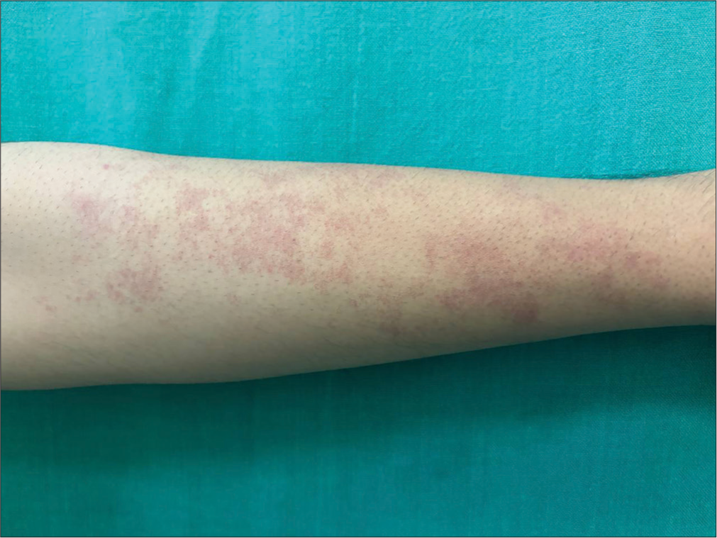
Angioma serpiginosum (AS) is a rare nevoid vascular disorder affecting the small vessels of the upper dermis. It is clinically characterized by multiple copper-red to red-colored grouped macules and papules during childhood. The lesions are usually unilateral with a linear serpiginous pattern following Blaschkoid distribution. It is rarely associated with extracutaneous findings. We present a 21-year-old male with multiple asymptomatic grouped pinpoint red lesions over the right arm for 6 years. These lesions initially appeared on the right upper arm and progressed to the forearm over a period of six years. There was no history of trauma, bleeding, or similar complaints in the family. General and systemic examination was normal. Cutaneous examination revealed multiple, punctate, grouped, bright red, and minimally raised papules over the right forearm, arranged linearly [Figure 1]. On Diascopy, the skin lesion was partially blanchable. Polarized dermoscopy using 3 Gen DermLite DL4 (CA, USA) ×10 magnification showed numerous small, grouped round to oval red dots and globules suggestive of lagoons [Figure 2]. Histopathological examination from an erythematous non-blanchable macule revealed the presence of multiple proliferating and dilated thin-walled capillaries with intraluminal presence of erythrocytes (thrombosed capillaries) in the papillary dermis with sparse perivascular lymphocytic infiltrate [Figures 3 and 4]. Hence based on clinical, dermoscopic, and histopathological features, a diagnosis of AS was made, and the patient was counseled about the condition and advised pulsed dye laser as it was of cosmetic concern to him. Hutchinson originally defined AS as a “serpiginous or infective nevus” in 1889.[1] Crocker later gave the condition its official name in 1894. The disorder is mostly sporadic; however, reports of family cases with an autosomal dominant inheritance have been made. AS affects people of all ages, but it is most common in childhood, with a 9:1 female-to-male ratio.[2] According to reports, the prevalence is <1:1,000,000.[2] The condition is usually asymptomatic, starts in early childhood, and stabilizes in adults. Although complete spontaneous involution is uncommon, partial involution is possible. There is still no established pathophysiology for AS. Elevated estrogen levels have been suggested as the etiology for the advancement of lesions during pregnancy and its prevalence in females; however, the lack of estrogen-progesterone receptors in the involved vessels in subsequent studies has disproved this theory. An additional theory is an aberrant vascular reaction to cold, which takes the form of newly formed big ectatic vasculature from developed capillaries in the dermal papillary layer. Mutation in the porcupine O-acyltransferase gene is often seen.[1,2] Clinically, lesions are copper to bright red, punctate, non-blanchable or partially blanchable, grouped macules that may develop into papules with background erythema. Lesions enlarge by developing new lesions at the periphery with the clearing of lesions in the center and this leads to a serpiginous or gyrate or ring-like morphology.[3]

- Multiple grouped pinpoint red macules and tiny papules over right forearm.
![Dermoscopic showing multiple clustered bright red lagoons with peripheral extensions (black circles) [DermLite 3 Gen DL 4, polarized ×10].](/content/130/2024/4/1/img/CSDM-4-141-g002.png)
- Dermoscopic showing multiple clustered bright red lagoons with peripheral extensions (black circles) [DermLite 3 Gen DL 4, polarized ×10].
![Scanner view of skin biopsy specimen showing dilated thin-walled capillaries in the papillary dermis [H&E, ×40]. H&E: Hematoxylin and eosin.](/content/130/2024/4/1/img/CSDM-4-141-g003.png)
- Scanner view of skin biopsy specimen showing dilated thin-walled capillaries in the papillary dermis [H&E, ×40]. H&E: Hematoxylin and eosin.
![Skin biopsy on higher magnification showing congested capillaries filled with red blood cells with sparse perivascular lymphocytic infiltrate [H&E, ×400]. H&E: Hematoxylin and eosin.](/content/130/2024/4/1/img/CSDM-4-141-g004.png)
- Skin biopsy on higher magnification showing congested capillaries filled with red blood cells with sparse perivascular lymphocytic infiltrate [H&E, ×400]. H&E: Hematoxylin and eosin.
Diascopy reveals non-blanchable to partially blanchable lesions. Dermoscopy is a valuable tool in diagnosing AS as it is a quick, non-invasive, and bedside aid that helps narrow down the differentials. It shows well-demarcated oval to round red lagoons (bright-red lacunae filled with blood) due to dilated vascular spaces within the papillary or superficial reticular dermis.[4] Histopathology reveals dilated thin-walled capillaries in the papillary dermis, lined by flattened endothelial cells of normal appearance.[4] AS needs to be differentiated from its close clinical mimickers that include unilateral nevoid telangiectasia, and pigmented purpuric dermatoses [Table 1].[5] Although the condition is limited to the skin, the ocular and nervous system may also be involved. Counseling and reassurance of this condition is of utmost importance as it may affect the self-esteem of the patient and treatment is indicated only for cosmetic reasons. Among the available treatment modalities cosmetic camouflage is an effective option and the treatment of choice is considered to be vascular lasers like pulsed dye lasers.[6] Other lasers mentioned in the literature include potassium titanyl phosphate lasers of 532 nm wavelength, intense pulsed light, Argon lasers, etc.[2]
| Angioma Serpiginosum | Unilateral Nevoid Telangiectasia | Pigmented Purpuric Dermatosis | |
|---|---|---|---|
| Introduction | A nevoid vascular disorder affecting the small vessels of the upper dermis | Rare congenital and acquired primary telangiectasia | Collection of generally benign, chronic, purpuric skin eruptions |
| Clinical Features | Multiple grouped, asymptomatic, bright red macules arranged in a linear manner along Blaschko’s lines | Multiple unilateral, linear, blanching telangiectases in a dermatomal or Blaschkoid pattern with an anemic halo around the lesion | Red to purple macules, patches and petechiae Most commonly occurs on lower extremities |
| Dermoscopy | Well-demarcated oval to round red lagoons | Red tortuous capillaries with a reticulated appearance | Coppery red to red-brown background, red dots and globules, dotted and blotchy red vessels in the perifollicular area |
| Histopathology | Dilated, thin-walled capillaries in papillary dermis | Multiple dilated, thin-walled vessels lined by plumped endothelial cells in the papillary and upper reticular dermis | Dilated capillaries in the dermis with extravasated RBC and hemosiderin pigment infiltrate |
In conclusion, AS is a rare entity in males, and in today’s era, dermoscopy acts as an essential bedside tool that sometimes can obviate the need for skin biopsy based on specific clues.
Ethical approval
The Institutional Review Board approval is not required.
Declaration of patient consent
The authors certify that they have obtained all appropriate patient consent.
Conflicts of interest
There are no conflicts of interest.
Use of artificial intelligence (AI)-assisted technology for manuscript preparation
The authors confirm that there was no use of artificial intelligence (AI)-assisted technology for assisting in the writing or editing of the manuscript and no images were manipulated using AI.
Financial support and sponsorship
Nil.
References
- Angioma serpiginosum: A case report and review of the literature. Ital J Pediatr. 2019;45:53.
- [CrossRef] [PubMed] [Google Scholar]
- Angioma serpiginosum In: StatPearls. Treasure Island, FL: Stat Pearls Publishing; 2022.
- [Google Scholar]
- Blaschkoid angioma serpiginosum: A dermoscopic diagnosis. Indian Dermatol Online J. 2018;9:127-9.
- [CrossRef] [PubMed] [Google Scholar]
- Dermoscopy of angioma serpiginosum. Indian J Dermatopathol Diagn Dermatol. 2022;9:41-3.
- [CrossRef] [Google Scholar]
- Angioma serpiginosum in a patchy and Blaschkoid distribution: A rare condition with an unconventional presentation. Indian J Dermatol. 2016;61:570-2.
- [CrossRef] [PubMed] [Google Scholar]


