
Translate this page into:
Barely visible yet intensely felt Itch: Dermoscopy clinches the diagnosis

*Corresponding author: Payal Chauhan, Department of Dermatology, All India Institute of Medical Sciences, Jammu, Jammu and Kashmir, India. chauhanpayal89@gmail.com
-
Received: ,
Accepted: ,
How to cite this article: Dhiman A, Daroach M, Chauhan P. Barely visible yet intensely felt Itch: Dermoscopy clinches the diagnosis. CosmoDerma. 2024;4:100. doi: 10.25259/CSDM_110_2024
Dear Sir,
A 68-year-old male, a known case of vitiligo vulgaris, presented with generalized intense itching for approximately two years. On examination, except from depigmented macules of vitiligo vulgaris, no lesions were evident [Figure 1]. The perplexing nature of pruritus prompted closer inspection with a dermoscope. When the dermoscope was put to use, pubic lice (Pthirus pubis) became apparent at multiple sites on the trunk [Figure 2]. Dermoscopy-assisted extraction of the mite was performed for further microscopic examination, which revealed an oval-shaped, broad structure with a small head, short antennae, three pairs of legs, the first pair being shorter and slender, and an abdominal region showing hairy processes known as setae, which confirmed crab lice infestation [Figure 3]. The patient was advised disinfection of clothes and bedding, along with topical permethrin 1% and oral ivermectin.
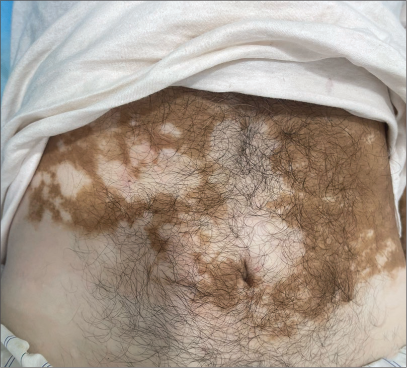
- No evident skin lesion apart from depigmented macules of vitiligo on initial examination.
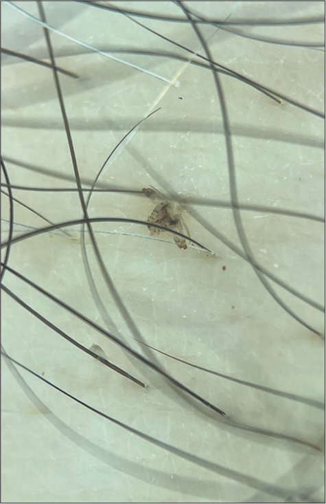
- Dermoscopy (DermLite DL5; 3Gen; San Juan Capistrano, California, USA, noncontact polarized mode, ×10 magnification, images capture with DermLite adapter for iPhone 12 pro max) demonstrated adult Pthirus pubis.
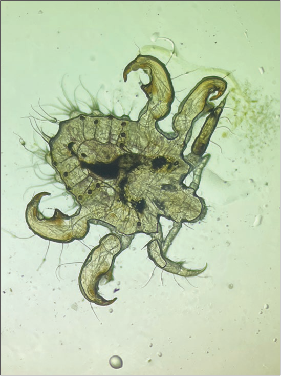
- Microscopic examination (Normal saline mount ×40) revealed an adult Pthirus pubis.
The P. pubis typically infests areas such as the pubic region, groin, buttocks, and perianal region. However, it may also affect other regions, especially in individuals with substantial body hair, including the thighs, abdomen, chest, underarms, and even beards. In certain cases, such as with children, it can even infest the eyelashes and eyebrows.[1]
Dermoscopy acted as a critical diagnostic tool in the present case of generalized pruritus sans visible skin lesions, by highlighting barely discernible small, pigmented dots on the patient’s skin to be crab lice, which might otherwise have gone unnoticed on routine naked eye examination. Moreover, dermoscopy also played an important role in the extraction of the imperceptible lice for microbiological examination and thus, clinching the diagnosis. Considering uncommon causes of generalized itching when standard clinical examination does not reveal any apparent lesions cannot be overstressed. To conclude, dermoscopy can act as a vital diagnostic modality for detecting subtle cutaneous manifestations of ectoparasitic infestations.
Ethical approval
The Institutional Review Board approval is not required.
Declaration of patient consent
The authors certify that they have obtained all appropriate patient consent.
Conflicts of interest
There are no conflicts of interest.
Use of artificial intelligence (AI)-assisted technology for manuscript preparation
The authors confirm that there was no use of artificial intelligence (AI)-assisted technology for assisting in the writing or editing of the manuscript and no images were manipulated using AI.
Financial support and sponsorship
Nil.
References
- Phthirus pubis infestation of the scalp: A case report and review of the literature. Korean J Parasitol. 2018;56:487-9.
- [CrossRef] [PubMed] [Google Scholar]


