
Translate this page into:
Medical illustration – Fusion of art, technology, and medical education


*Corresponding author: Yogesh Ashok Sontakke, Department of Anatomy, JIPMER, Academic Center, Pondicherry, India. dryogeshas@gmail.com
-
Received: ,
Accepted: ,
How to cite this article: Sontakke YA. Medical illustration – Fusion of art, technology, and medical education. CosmoDerma 2022;2:121.
INTRODUCTION
An illustration is a visual representation of a text that decoratively depicts the facts or description. The illustration eases the readability of the text. Most medical facts can be easily explained using medical illustrations rather than describing them in words. It represents an idea of a presenter in the form of images, flowcharts, models, or animations, and so on.[1,2]
The medical illustration has a long history. Aristotle (384–322 BC) used papyrus scrolls to write his pictorial works. Leonardo da Vinci (1452–1519) produced 750 drawings of cadavers. Andreas Vesalius wrote a series of seven books, De Humani Corporis Fabrica Libri, in 1543. His drawings are historically displayed on the walls of most medical schools worldwide. Frank Henry Netter (1906–1991) produced nearly 4000 illustrations using gouache paints and illustration boards.[2,3] Over a period, the journey of medical illustrations has been revolutionized due to the advancement of printing media, computers, digital drawing software, and hardware. The field of medical illustration is wide and interesting. It gives the opportunity to communicate effectively. Scientific knowledge is essential for creating a medical illustration.
USES OF MEDICAL ILLUSTRATIONS
There are numerous uses of medical illustrations, a few of them are as follows:
Publications of textbook, journals, and e-books
Web content
Patient education
Medical education
Interactive learning
Cell phone health applications
Museums
Film and television
Augmented and virtual reality.
ADVANTAGES AND DISADVANTAGES OF MEDICAL ILLUSTRATIONS
For each fact, there are always advantages and disadvantages.
Advantages of medical illustrations
They save time in explaining the desired fact
They are an effective way of communication
They can be preserved in electronic media for a longer time and can be reproduced as per the need
They are suitable for transmission through electronic media to a wider audience.
Disadvantages of medical illustrations
The creation of a good illustration consumes lot of time
Illustrations can never replace clinical patient examination
Illustrations cannot replace practical procedures such as suturing a wound
Creation of illustration requires knowledge of art and software
Cost for professional medical illustration may be high. The software may need subscription cost.
TYPES OF MEDICAL ILLUSTRATIONS
The medical illustrations can be grouped as narrative, decorative, informative, and conceptual.
Narrative illustration
These give an idea about the entire book or process. These are useful as the cover page of a book, medical magazine, or main illustration of a brochure or website. It is an umbrella of the entire topic.
Decorative illustration
These utilize designs to simplify or exaggerate the topic. They are more useful for attracting the attention of the audience or reader. They are made in a less scientific manner, for example, affected ugly part of a beautiful face to show cancerous changes in smokers.
Informative illustration
These utilize the facts to be represented. These are more scientific and the most used in textbooks and scientific literature. They summarize facts that require space and create a message.
Conceptual illustration
These are created from an idea over the content. The images are created using reality to simplify the subject, for example, line diagrams of dissected cadaveric parts.
All the groups mentioned above have overlapping domains. To classify, an illustration in a specific group may sometimes be difficult. Illustration includes photographs, drawings, diagrams, and flowcharts. They may be black and white, colorful, two-dimensional, or three-dimensional.
PROCESS OF CREATION OF MEDICAL ILLUSTRATION
Pre-concept factors
The factors to be considered for medical illustration [Figure 1]:
Purpose of illustration
Audience or readers for whom it is created
Mode of display such as print media, painting, electronic media, computer- or cell-phone-based applications, and so on
Selection of colors: The colors should be selected carefully to depict the human body structures scientifically.

- Planning of medical illustration.
Selection of a topic
For drawing the medical illustration, selecting the topic and understanding the need is important. It involves the following components [Figure 1]:
The topic should be read carefully with pre-topics and post-topics that are covered. For example, showing the passage of contents of an indirect inguinal hernia, the topic can be supported with an illustration of inguinal canal, and further, it may be followed by illustration showing surgical correction of the hernia.
Audience: Before starting the drawing, the audience should be known. For example, illustrations for postgraduates will be more complex than for undergrads.
Color scheme: The color scheme has scientific coding that needs to be followed. For example, RBCs in red color can be used. Similarly, to show RBCs with oxygenated hemoglobin bright red and with deoxygenated hemoglobin, blue (purple) color can be used. Follow the same color scheme throughout the entire document.
Selection of type of illustration
Suitable type of illustration needs to be selected based on the topic, use (cover page, poster, and webpage), audience, and data. The illustration can be raster or vector. The examples of two types of illustrations derived from the same text are shown in [Figure 1].
Selection of software
The raster illustrations can be drawn using Adobe Photoshop, MacPaint, PC paintbrush, painted, and so on. The vector illustrations can be drawn using Adobe illustration, free hand, MacDraw, Expression, and so on. Software such as canvas and CorelDraw are useful for creating raster and vector images. All these software have pros and cons over each other.[4-7]
Sketching and plotting
Based on the points mentioned above, rough sketching can be prepared.
Drawing illustration
Using suitable software, rough sketch is converted into a final illustration. During drawing the illustration, the focus should be kept on the following points [Figure 2–5]:
Three-dimensional effects
Line, shape, tone, color, scale, and empty space to enhance the effects.
Space of illustration is divided into positive space which is occupied by the illustration, and negative space which is unoccupied space surrounding the illustration. Both have their significance in making illustration effective.
Text in illustration should have proper font style, readable font size, and suitable contrast, shadow, and texture to elevate the illustration.
Simplicity in the illustration makes it suitable for most of the audience.
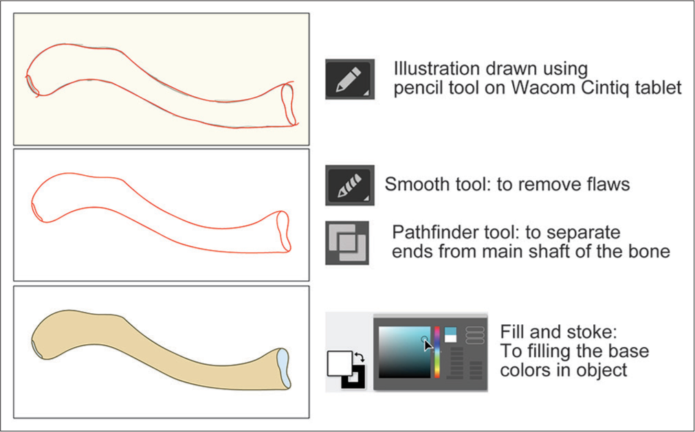
- Drawing medical illustration using illustrator software.

- Adding details in illustration.

- Example for preparation of medical illustration. (a) Text selected for the preparation of medical illustration. (b) Schematic representation of the text. (c) Flowchart representation of text.

- Another example for preparation of medical illustration. (a) Text selected for the preparation of medical illustration. (b) Diagrammatic representation of the text (Image courtesy: Textbook of Human Histology, Yogesh Sontakke, 1st Edn. CBSPD, Delhi, India).
Review and feedback
To obtain the proper feedback, give the illustration to colleagues and few participants from the audience group. Check the perfectness of the transmission of the intended message. Obtain both positive and negative feedback. Analyze the feedback carefully and modify the illustration saving the original one. Repeat the process of feedback until a satisfactory result is obtained. Always keep in mind the illustration is for audience, and they should understand the given message. The illustration should depict the written text. It should not be ambiguous.
ETHICS AND COPYRIGHT
The medical illustration should not be misleading or unnecessarily exaggerating the facts and should not hurt the emotions of the audience. It should not defame or convey the wrong message to the audience. Care should be taken while using patient photographs. The individual’s identity should never be disclosed, and it can be masked whenever possible and should be published with written consent from the patient.[8] Do not copy the illustrations; recreate them with inputs of new ideas. Copyrighted illustrations should be published only after obtaining written permission from the owner, who may be a publisher or artist.
CONCLUSION
Medical illustrations and the art behind them are an integral part of medical education. Various factors need to be considered while creating the illustration, ranging from pencil drawing to software-based image creation, for public awareness to undergraduate teaching.
Declaration of patient consent
Patient’s consent not required as there are no patients in this study.
Financial support and sponsorship
Nil.
Conflicts of interest
There are no conflicts of interest.
References
- Changing art of anatomy illustrations. Sch Int J Anat Physiol. 2022;5:55-8.
- [CrossRef] [Google Scholar]
- Medical illustration: Art in medical education. Heart Views. 2011;12:83-91.
- [CrossRef] [PubMed] [Google Scholar]
- Medical illustration: From caves to cyberspace. Health Info Libr J. 2001;18:99-109.
- [CrossRef] [PubMed] [Google Scholar]
- Preparing effective medical illustrations for publication (Part 2): Software processing, drawing and illustration. Biomed Imaging Interv J. 2008;4:e12.
- [CrossRef] [Google Scholar]
- Digital medical illustration for the radiologist. Radiographics. 2018;38:1145-57.
- [CrossRef] [PubMed] [Google Scholar]
- Digital art-a useful tool for medical professionals to create medical illustrations. JPRAS Open. 2021;28:97-102.
- [CrossRef] [PubMed] [Google Scholar]
- Collaborating with medical illustrators to create optimal surgical figures for publications and beyond. J Plast Reconstr Aesthet Surg. 2022;75:2001-8.
- [CrossRef] [PubMed] [Google Scholar]
- Ethical and legal aspects on the use of images and photographs in medical teaching and publication. Int Angiol. 2010;29:376-9.
- [Google Scholar]


