Generic selectors
Exact matches only
Search in title
Search in content
Post Type Selectors
Filter by Categories
Brief Report
Case Report
Editorial
Focus
Images/Instrument in Dermatology/Dermatosurgery
Innovations
Letter to the Editor
Living Legends
Looking back in history
Original Article
Perspective
Resident Forum
Review Article
Spot the Diagnosis
Tropical Dermatology
Visual Treats in Dermatology
View/Download PDF

Translate this page into:

Visual Treats in Dermatology
2022
:2;
2
doi:
10.25259/CSDM_71_2021

Subcutaneous zygomycosis

Department of Dermatology, Jawaharlal Institute of Postgraduate Medical Education and Research, Puducherry, India
Corresponding author: Arunachalam Narayanan, Department of Dermatology, Jawaharlal Institute of Postgraduate Medical Education and Research, Puducherry, India. narayanan359@gmail.com
Licence
This is an open-access article distributed under the terms of the Creative Commons Attribution-Non Commercial-Share Alike 4.0 License, which allows others to remix, transform, and build upon the work non-commercially, as long as the author is credited and the new creations are licensed under the identical terms.

How to cite this article: Narayanan A, Anurag SV. Subcutaneous zygomycosis. CosmoDerma 2022;2:2.

A 55-year-old male patient presented with a painless, subcutaneous, slowly spreading, disc-shaped, indurated plaque over the anterolateral aspect of the left thigh [Figure 1] for 6 months. Fingers could be insinuated under the disc-like plaque from the periphery of the lesion. There was no history of prior trauma, pain, tenderness, itching, warmth, erythema, or discharge from the plaque. Histopathological examination revealed mixed inflammatory infiltrate in the subcutis with few scattered aseptate fungal hyphae surrounded by eosinophilic material suggestive of Splendore-Hoeppli phenomenon. Based on these clinical and histopathological findings, we made a diagnosis of subcutaneous zygomycosis. Subcutaneous zygomycosis caused by Basidiobolus ranarum typically involves the thighs and buttocks. The infection nearly almost always occurs in immunocompetent individuals.[1] Treatment options include systemic therapy with potassium iodide, itraconazole, amphotericin B, local hyperbaric oxygen, and surgical debridement.

Figure 1:
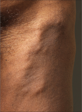
Painless, subcutaneous, slowly spreading, disc-shaped, indurated plaque over the anterolateral aspect of the left thigh.

Declaration of patient consent

Patient’s consent is not required as patient’s identity is neither disclosed nor compromised.

Financial support and sponsorship

Nil.

Conflicts of interest

There are no conflicts of interest.

References

  1. , , , , , . Subcutaneous zygomycosis: Report of 10 cases from two institutions in North India. J Eur Acad Dermatol Venereol. 2010;24:1220-5.
    [CrossRef] [PubMed] [Google Scholar]

Fulltext Views
4,334

PDF downloads
475
View/Download PDF
Download Citations
BibTeX
RIS
Show Sections